Chlorine »
PDB 3k9u-3khi »
3keb »
Chlorine in PDB 3keb: Thiol Peroxidase From Chromobacterium Violaceum
Protein crystallography data
The structure of Thiol Peroxidase From Chromobacterium Violaceum, PDB code: 3keb
was solved by
J.Osipiuk,
O.Kagan,
A.Savchenko,
A.M.Edwards,
A.Joachimiak,
Midwest Centerfor Structural Genomics (Mcsg),
with X-Ray Crystallography technique. A brief refinement statistics is given in the table below:
| Resolution Low / High (Å) | 43.00 / 1.80 |
| Space group | P 1 21 1 |
| Cell size a, b, c (Å), α, β, γ (°) | 74.630, 59.387, 93.484, 90.00, 98.30, 90.00 |
| R / Rfree (%) | 20.6 / 24.2 |
Chlorine Binding Sites:
The binding sites of Chlorine atom in the Thiol Peroxidase From Chromobacterium Violaceum
(pdb code 3keb). This binding sites where shown within
5.0 Angstroms radius around Chlorine atom.
In total 3 binding sites of Chlorine where determined in the Thiol Peroxidase From Chromobacterium Violaceum, PDB code: 3keb:
Jump to Chlorine binding site number: 1; 2; 3;
In total 3 binding sites of Chlorine where determined in the Thiol Peroxidase From Chromobacterium Violaceum, PDB code: 3keb:
Jump to Chlorine binding site number: 1; 2; 3;
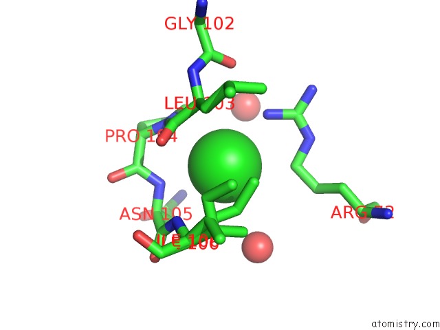
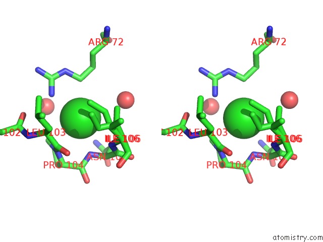
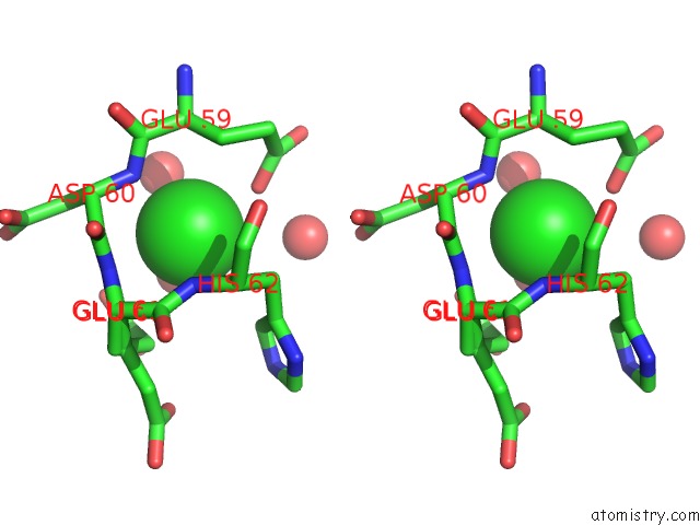
Chlorine binding site 1 out of 3 in 3keb
Go back to
Chlorine binding site 1 out
of 3 in the Thiol Peroxidase From Chromobacterium Violaceum
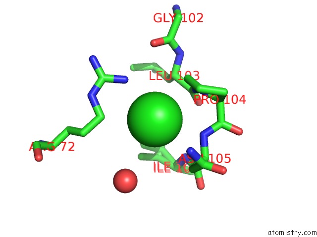
Mono view

Stereo pair view
Mono view
Stereo pair view
A full contact list of Chlorine with other atoms in the Cl binding
site number 1 of Thiol Peroxidase From Chromobacterium Violaceum within 5.0Å range:
|
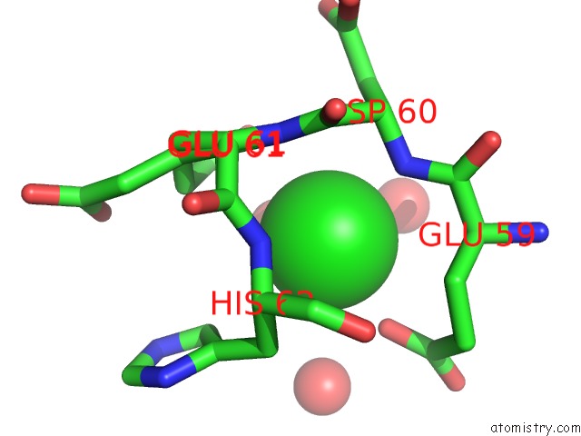
Chlorine binding site 2 out of 3 in 3keb
Go back to
Chlorine binding site 2 out
of 3 in the Thiol Peroxidase From Chromobacterium Violaceum
Mono view
Stereo pair view
Mono view
Stereo pair view
A full contact list of Chlorine with other atoms in the Cl binding
site number 2 of Thiol Peroxidase From Chromobacterium Violaceum within 5.0Å range:
|
Chlorine binding site 3 out of 3 in 3keb
Go back to
Chlorine binding site 3 out
of 3 in the Thiol Peroxidase From Chromobacterium Violaceum
Mono view
Stereo pair view
Mono view
Stereo pair view
A full contact list of Chlorine with other atoms in the Cl binding
site number 3 of Thiol Peroxidase From Chromobacterium Violaceum within 5.0Å range:
|
Reference:
J.Osipiuk,
O.Kagan,
A.Savchenko,
A.M.Edwards,
A.Joachimiak.
X-Ray Crystal Structure of Thiol Peroxidase From Chromobacterium Violaceum To Be Published.
Page generated: Sat Jul 20 22:36:30 2024
Last articles
Ca in 5SWICa in 5SVE
Ca in 5SSX
Ca in 5SV0
Ca in 5STD
Ca in 5SSZ
Ca in 5SSY
Ca in 5SIC
Ca in 5SBD
Ca in 5SBE