Chlorine »
PDB 3khp-3ktf »
3kia »
Chlorine in PDB 3kia: Crystal Structure of Mannosyl-3-Phosphoglycerate Synthase From Rubrobacter Xylanophilus
Enzymatic activity of Crystal Structure of Mannosyl-3-Phosphoglycerate Synthase From Rubrobacter Xylanophilus
All present enzymatic activity of Crystal Structure of Mannosyl-3-Phosphoglycerate Synthase From Rubrobacter Xylanophilus:
2.4.1.217;
2.4.1.217;
Protein crystallography data
The structure of Crystal Structure of Mannosyl-3-Phosphoglycerate Synthase From Rubrobacter Xylanophilus, PDB code: 3kia
was solved by
S.Macedo-Ribeiro,
P.J.B.Pereira,
N.Empadinhas,
M.S.Da Costa,
with X-Ray Crystallography technique. A brief refinement statistics is given in the table below:
| Resolution Low / High (Å) | 53.70 / 2.80 |
| Space group | P 65 2 2 |
| Cell size a, b, c (Å), α, β, γ (°) | 109.010, 109.010, 313.418, 90.00, 90.00, 120.00 |
| R / Rfree (%) | 19 / 23.9 |
Other elements in 3kia:
The structure of Crystal Structure of Mannosyl-3-Phosphoglycerate Synthase From Rubrobacter Xylanophilus also contains other interesting chemical elements:
| Magnesium | (Mg) | 3 atoms |
Chlorine Binding Sites:
The binding sites of Chlorine atom in the Crystal Structure of Mannosyl-3-Phosphoglycerate Synthase From Rubrobacter Xylanophilus
(pdb code 3kia). This binding sites where shown within
5.0 Angstroms radius around Chlorine atom.
In total 3 binding sites of Chlorine where determined in the Crystal Structure of Mannosyl-3-Phosphoglycerate Synthase From Rubrobacter Xylanophilus, PDB code: 3kia:
Jump to Chlorine binding site number: 1; 2; 3;
In total 3 binding sites of Chlorine where determined in the Crystal Structure of Mannosyl-3-Phosphoglycerate Synthase From Rubrobacter Xylanophilus, PDB code: 3kia:
Jump to Chlorine binding site number: 1; 2; 3;
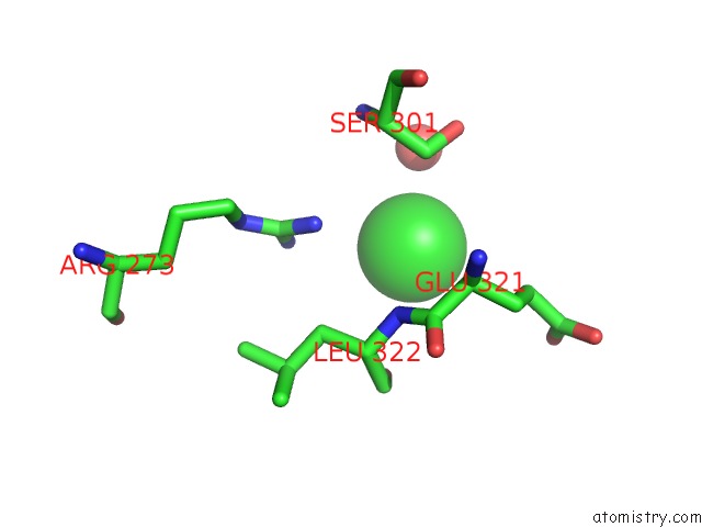
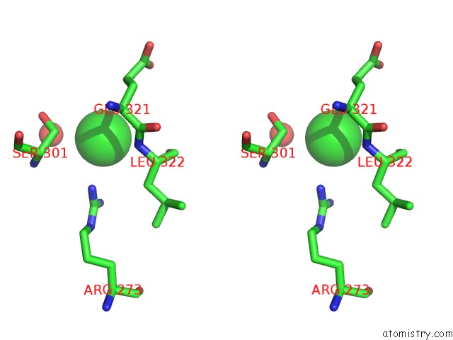
Chlorine binding site 1 out of 3 in 3kia
Go back to
Chlorine binding site 1 out
of 3 in the Crystal Structure of Mannosyl-3-Phosphoglycerate Synthase From Rubrobacter Xylanophilus
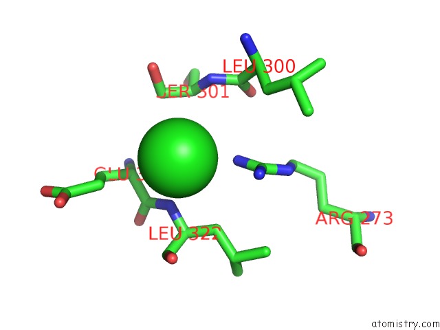
Mono view
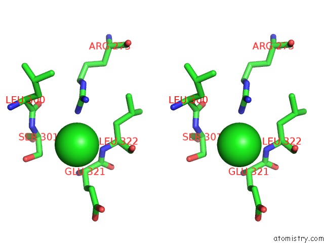
Stereo pair view
Mono view
Stereo pair view
A full contact list of Chlorine with other atoms in the Cl binding
site number 1 of Crystal Structure of Mannosyl-3-Phosphoglycerate Synthase From Rubrobacter Xylanophilus within 5.0Å range:
|
Chlorine binding site 2 out of 3 in 3kia
Go back to
Chlorine binding site 2 out
of 3 in the Crystal Structure of Mannosyl-3-Phosphoglycerate Synthase From Rubrobacter Xylanophilus
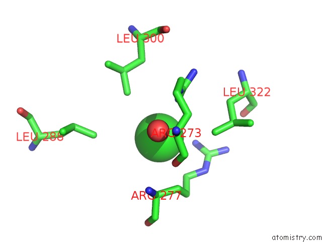
Mono view
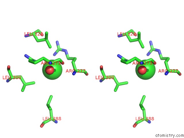
Stereo pair view
Mono view
Stereo pair view
A full contact list of Chlorine with other atoms in the Cl binding
site number 2 of Crystal Structure of Mannosyl-3-Phosphoglycerate Synthase From Rubrobacter Xylanophilus within 5.0Å range:
|
Chlorine binding site 3 out of 3 in 3kia
Go back to
Chlorine binding site 3 out
of 3 in the Crystal Structure of Mannosyl-3-Phosphoglycerate Synthase From Rubrobacter Xylanophilus
Mono view
Stereo pair view
Mono view
Stereo pair view
A full contact list of Chlorine with other atoms in the Cl binding
site number 3 of Crystal Structure of Mannosyl-3-Phosphoglycerate Synthase From Rubrobacter Xylanophilus within 5.0Å range:
|
Reference:
N.Empadinhas,
P.J.B.Pereira,
L.Albuquerque,
J.Costa,
B.Sa-Moura,
A.T.Marques,
S.Macedo-Ribeiro,
M.S.Da Costa.
Functional and Structural Characterization of A Novel Mannosyl-3-Phosphoglycerate Synthase From Rubrobacter Xylanophilus Reveals Its Dual Substrate Specificity Mol.Microbiol. V. 79 76 2011.
ISSN: ISSN 0950-382X
PubMed: 21166895
DOI: 10.1111/J.1365-2958.2010.07432.X
Page generated: Fri Jul 11 07:00:35 2025
ISSN: ISSN 0950-382X
PubMed: 21166895
DOI: 10.1111/J.1365-2958.2010.07432.X
Last articles
Fe in 2YXOFe in 2YRS
Fe in 2YXC
Fe in 2YNM
Fe in 2YVJ
Fe in 2YP1
Fe in 2YU2
Fe in 2YU1
Fe in 2YQB
Fe in 2YOO