Chlorine »
PDB 3khp-3ktf »
3koo »
Chlorine in PDB 3koo: Crystal Structure of Soluble Epoxide Hydrolase
Enzymatic activity of Crystal Structure of Soluble Epoxide Hydrolase
All present enzymatic activity of Crystal Structure of Soluble Epoxide Hydrolase:
3.3.2.10;
3.3.2.10;
Protein crystallography data
The structure of Crystal Structure of Soluble Epoxide Hydrolase, PDB code: 3koo
was solved by
N.A.Farrow,
with X-Ray Crystallography technique. A brief refinement statistics is given in the table below:
| Resolution Low / High (Å) | 48.20 / 2.79 |
| Space group | P 65 2 2 |
| Cell size a, b, c (Å), α, β, γ (°) | 91.765, 91.765, 242.516, 90.00, 90.00, 120.00 |
| R / Rfree (%) | 21.3 / 30.7 |
Chlorine Binding Sites:
The binding sites of Chlorine atom in the Crystal Structure of Soluble Epoxide Hydrolase
(pdb code 3koo). This binding sites where shown within
5.0 Angstroms radius around Chlorine atom.
In total 2 binding sites of Chlorine where determined in the Crystal Structure of Soluble Epoxide Hydrolase, PDB code: 3koo:
Jump to Chlorine binding site number: 1; 2;
In total 2 binding sites of Chlorine where determined in the Crystal Structure of Soluble Epoxide Hydrolase, PDB code: 3koo:
Jump to Chlorine binding site number: 1; 2;
Chlorine binding site 1 out of 2 in 3koo
Go back to
Chlorine binding site 1 out
of 2 in the Crystal Structure of Soluble Epoxide Hydrolase
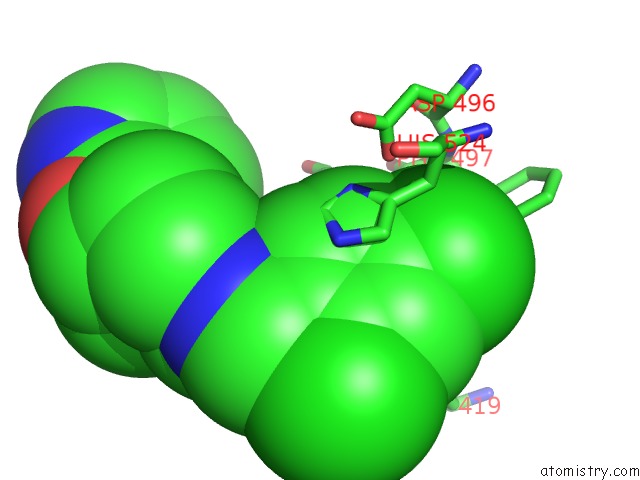
Mono view
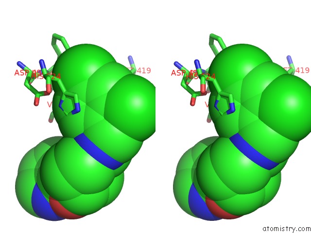
Stereo pair view
Mono view
Stereo pair view
A full contact list of Chlorine with other atoms in the Cl binding
site number 1 of Crystal Structure of Soluble Epoxide Hydrolase within 5.0Å range:
|
Chlorine binding site 2 out of 2 in 3koo
Go back to
Chlorine binding site 2 out
of 2 in the Crystal Structure of Soluble Epoxide Hydrolase
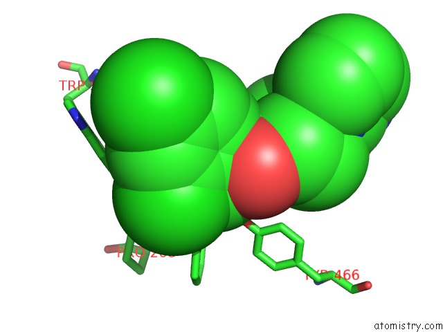
Mono view
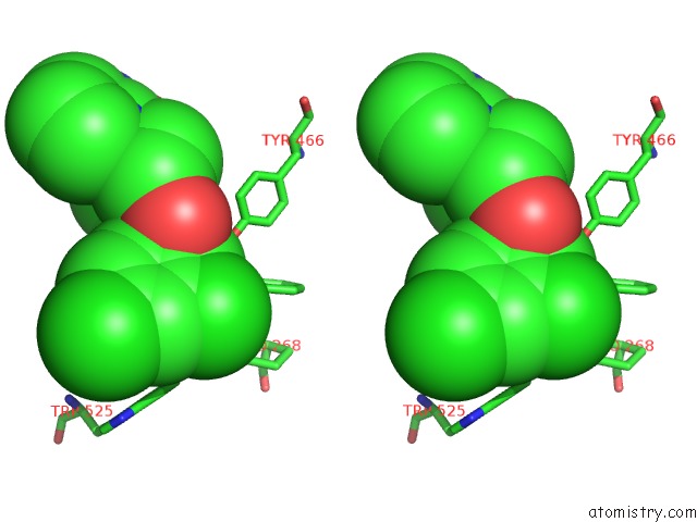
Stereo pair view
Mono view
Stereo pair view
A full contact list of Chlorine with other atoms in the Cl binding
site number 2 of Crystal Structure of Soluble Epoxide Hydrolase within 5.0Å range:
|
Reference:
A.B.Eldrup,
F.Soleymanzadeh,
N.A.Farrow,
A.Kukulka,
S.De Lombaert.
Optimization of Piperidyl-Ureas As Inhibitors of Soluble Epoxide Hydrolase. Bioorg.Med.Chem.Lett. V. 20 571 2010.
ISSN: ISSN 0960-894X
PubMed: 19969453
DOI: 10.1016/J.BMCL.2009.11.091
Page generated: Sat Jul 20 22:45:36 2024
ISSN: ISSN 0960-894X
PubMed: 19969453
DOI: 10.1016/J.BMCL.2009.11.091
Last articles
Zn in 9J0NZn in 9J0O
Zn in 9J0P
Zn in 9FJX
Zn in 9EKB
Zn in 9C0F
Zn in 9CAH
Zn in 9CH0
Zn in 9CH3
Zn in 9CH1