Chlorine »
PDB 3ku9-3l29 »
3l12 »
Chlorine in PDB 3l12: Crystal Structure of Putative Glycerophosphoryl Diester Phosphodiesterase (YP_165505.1) From Silicibacter Pomeroyi Dss-3 at 1.60 A Resolution
Protein crystallography data
The structure of Crystal Structure of Putative Glycerophosphoryl Diester Phosphodiesterase (YP_165505.1) From Silicibacter Pomeroyi Dss-3 at 1.60 A Resolution, PDB code: 3l12
was solved by
Joint Center For Structural Genomics (Jcsg),
with X-Ray Crystallography technique. A brief refinement statistics is given in the table below:
| Resolution Low / High (Å) | 27.28 / 1.60 |
| Space group | P 1 21 1 |
| Cell size a, b, c (Å), α, β, γ (°) | 50.409, 136.411, 50.548, 90.00, 118.00, 90.00 |
| R / Rfree (%) | 15.6 / 18.7 |
Other elements in 3l12:
The structure of Crystal Structure of Putative Glycerophosphoryl Diester Phosphodiesterase (YP_165505.1) From Silicibacter Pomeroyi Dss-3 at 1.60 A Resolution also contains other interesting chemical elements:
| Magnesium | (Mg) | 3 atoms |
Chlorine Binding Sites:
The binding sites of Chlorine atom in the Crystal Structure of Putative Glycerophosphoryl Diester Phosphodiesterase (YP_165505.1) From Silicibacter Pomeroyi Dss-3 at 1.60 A Resolution
(pdb code 3l12). This binding sites where shown within
5.0 Angstroms radius around Chlorine atom.
In total 5 binding sites of Chlorine where determined in the Crystal Structure of Putative Glycerophosphoryl Diester Phosphodiesterase (YP_165505.1) From Silicibacter Pomeroyi Dss-3 at 1.60 A Resolution, PDB code: 3l12:
Jump to Chlorine binding site number: 1; 2; 3; 4; 5;
In total 5 binding sites of Chlorine where determined in the Crystal Structure of Putative Glycerophosphoryl Diester Phosphodiesterase (YP_165505.1) From Silicibacter Pomeroyi Dss-3 at 1.60 A Resolution, PDB code: 3l12:
Jump to Chlorine binding site number: 1; 2; 3; 4; 5;
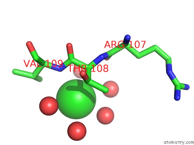
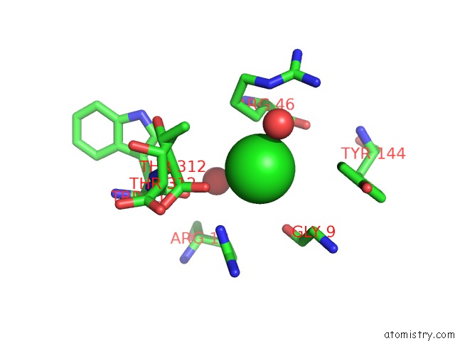
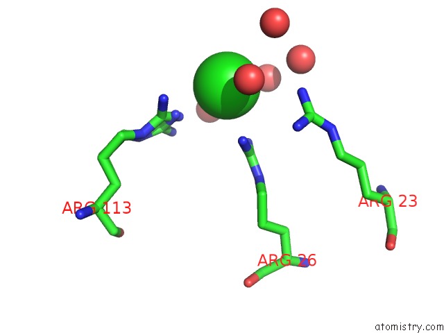
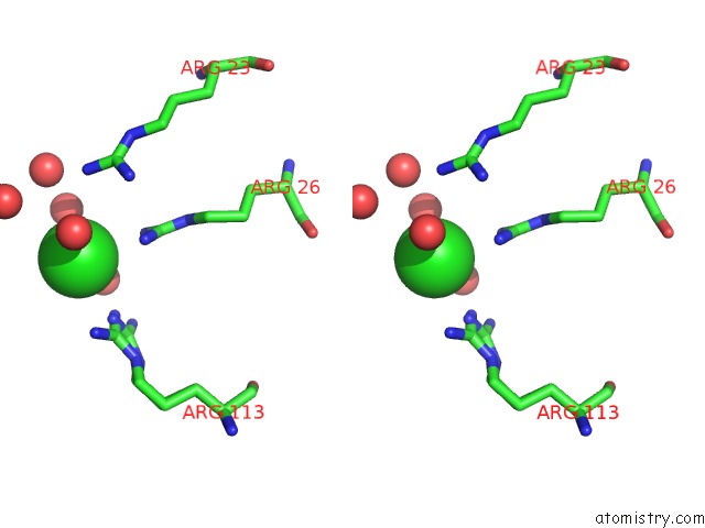
Chlorine binding site 1 out of 5 in 3l12
Go back to
Chlorine binding site 1 out
of 5 in the Crystal Structure of Putative Glycerophosphoryl Diester Phosphodiesterase (YP_165505.1) From Silicibacter Pomeroyi Dss-3 at 1.60 A Resolution
Mono view
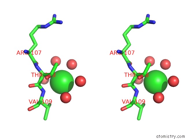
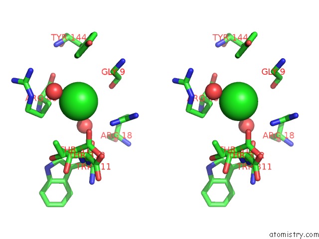
Stereo pair view
Mono view
Stereo pair view
A full contact list of Chlorine with other atoms in the Cl binding
site number 1 of Crystal Structure of Putative Glycerophosphoryl Diester Phosphodiesterase (YP_165505.1) From Silicibacter Pomeroyi Dss-3 at 1.60 A Resolution within 5.0Å range:
|
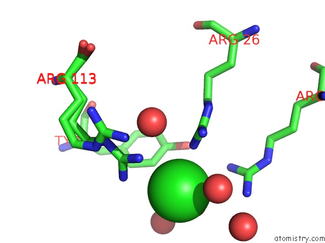
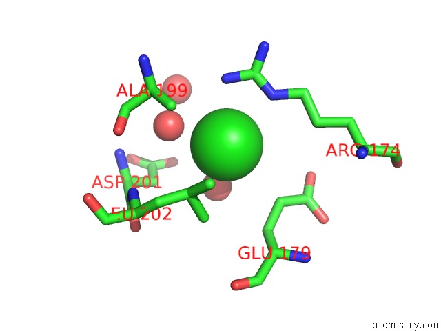
Chlorine binding site 2 out of 5 in 3l12
Go back to
Chlorine binding site 2 out
of 5 in the Crystal Structure of Putative Glycerophosphoryl Diester Phosphodiesterase (YP_165505.1) From Silicibacter Pomeroyi Dss-3 at 1.60 A Resolution
Mono view
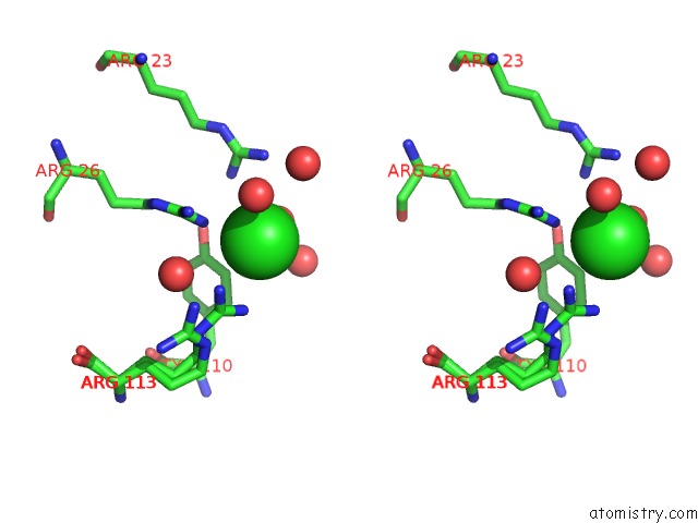
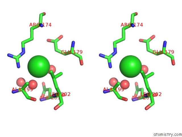
Stereo pair view
Mono view
Stereo pair view
A full contact list of Chlorine with other atoms in the Cl binding
site number 2 of Crystal Structure of Putative Glycerophosphoryl Diester Phosphodiesterase (YP_165505.1) From Silicibacter Pomeroyi Dss-3 at 1.60 A Resolution within 5.0Å range:
|
Chlorine binding site 3 out of 5 in 3l12
Go back to
Chlorine binding site 3 out
of 5 in the Crystal Structure of Putative Glycerophosphoryl Diester Phosphodiesterase (YP_165505.1) From Silicibacter Pomeroyi Dss-3 at 1.60 A Resolution
Mono view
Stereo pair view
Mono view
Stereo pair view
A full contact list of Chlorine with other atoms in the Cl binding
site number 3 of Crystal Structure of Putative Glycerophosphoryl Diester Phosphodiesterase (YP_165505.1) From Silicibacter Pomeroyi Dss-3 at 1.60 A Resolution within 5.0Å range:
|
Chlorine binding site 4 out of 5 in 3l12
Go back to
Chlorine binding site 4 out
of 5 in the Crystal Structure of Putative Glycerophosphoryl Diester Phosphodiesterase (YP_165505.1) From Silicibacter Pomeroyi Dss-3 at 1.60 A Resolution
Mono view
Stereo pair view
Mono view
Stereo pair view
A full contact list of Chlorine with other atoms in the Cl binding
site number 4 of Crystal Structure of Putative Glycerophosphoryl Diester Phosphodiesterase (YP_165505.1) From Silicibacter Pomeroyi Dss-3 at 1.60 A Resolution within 5.0Å range:
|
Chlorine binding site 5 out of 5 in 3l12
Go back to
Chlorine binding site 5 out
of 5 in the Crystal Structure of Putative Glycerophosphoryl Diester Phosphodiesterase (YP_165505.1) From Silicibacter Pomeroyi Dss-3 at 1.60 A Resolution
Mono view
Stereo pair view
Mono view
Stereo pair view
A full contact list of Chlorine with other atoms in the Cl binding
site number 5 of Crystal Structure of Putative Glycerophosphoryl Diester Phosphodiesterase (YP_165505.1) From Silicibacter Pomeroyi Dss-3 at 1.60 A Resolution within 5.0Å range:
|
Reference:
Joint Center For Structural Genomics (Jcsg),
Joint Center For Structural Genomics (Jcsg).
N/A N/A.
Page generated: Fri Jul 11 07:10:59 2025
Last articles
Cl in 5TZOCl in 5U09
Cl in 5TY2
Cl in 5TZL
Cl in 5TYK
Cl in 5TWS
Cl in 5TXU
Cl in 5TXI
Cl in 5TWR
Cl in 5TWO