Chlorine »
PDB 3l2h-3lcc »
3l6t »
Chlorine in PDB 3l6t: Crystal Structure of An N-Terminal Mutant of the Plasmid PCU1 Trai Relaxase Domain
Protein crystallography data
The structure of Crystal Structure of An N-Terminal Mutant of the Plasmid PCU1 Trai Relaxase Domain, PDB code: 3l6t
was solved by
M.R.Redinbo,
R.P.Nash,
with X-Ray Crystallography technique. A brief refinement statistics is given in the table below:
| Resolution Low / High (Å) | 31.37 / 1.93 |
| Space group | P 1 21 1 |
| Cell size a, b, c (Å), α, β, γ (°) | 50.385, 58.656, 86.532, 90.00, 95.01, 90.00 |
| R / Rfree (%) | 16.8 / 20.4 |
Other elements in 3l6t:
The structure of Crystal Structure of An N-Terminal Mutant of the Plasmid PCU1 Trai Relaxase Domain also contains other interesting chemical elements:
| Nickel | (Ni) | 2 atoms |
| Magnesium | (Mg) | 1 atom |
| Sodium | (Na) | 1 atom |
Chlorine Binding Sites:
The binding sites of Chlorine atom in the Crystal Structure of An N-Terminal Mutant of the Plasmid PCU1 Trai Relaxase Domain
(pdb code 3l6t). This binding sites where shown within
5.0 Angstroms radius around Chlorine atom.
In total 3 binding sites of Chlorine where determined in the Crystal Structure of An N-Terminal Mutant of the Plasmid PCU1 Trai Relaxase Domain, PDB code: 3l6t:
Jump to Chlorine binding site number: 1; 2; 3;
In total 3 binding sites of Chlorine where determined in the Crystal Structure of An N-Terminal Mutant of the Plasmid PCU1 Trai Relaxase Domain, PDB code: 3l6t:
Jump to Chlorine binding site number: 1; 2; 3;
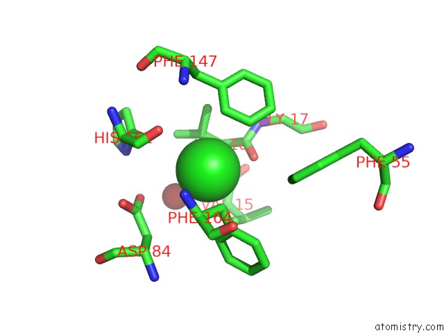
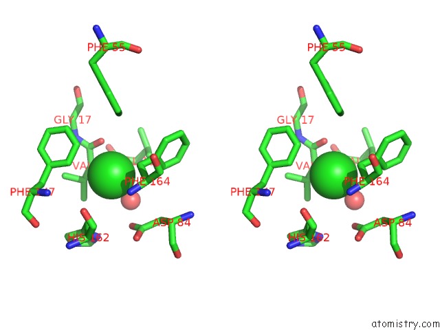
Chlorine binding site 1 out of 3 in 3l6t
Go back to
Chlorine binding site 1 out
of 3 in the Crystal Structure of An N-Terminal Mutant of the Plasmid PCU1 Trai Relaxase Domain
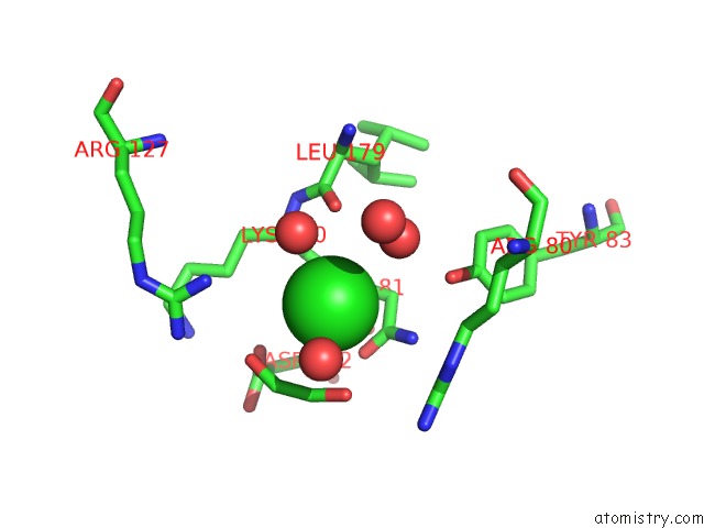
Mono view
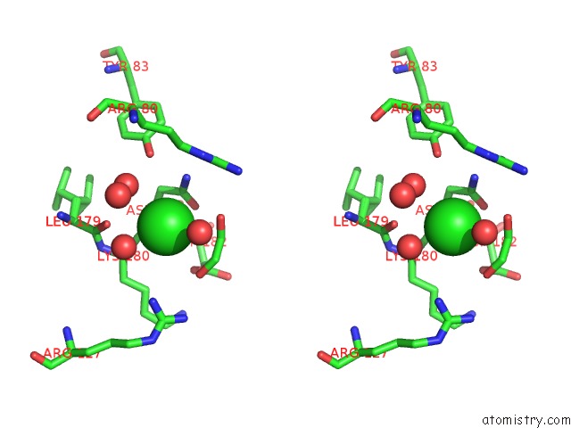
Stereo pair view
Mono view
Stereo pair view
A full contact list of Chlorine with other atoms in the Cl binding
site number 1 of Crystal Structure of An N-Terminal Mutant of the Plasmid PCU1 Trai Relaxase Domain within 5.0Å range:
|
Chlorine binding site 2 out of 3 in 3l6t
Go back to
Chlorine binding site 2 out
of 3 in the Crystal Structure of An N-Terminal Mutant of the Plasmid PCU1 Trai Relaxase Domain
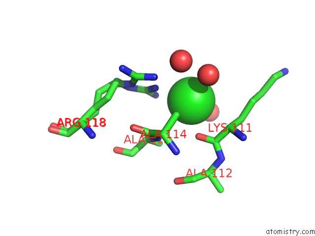
Mono view
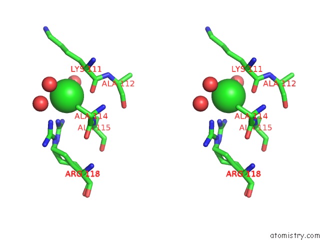
Stereo pair view
Mono view
Stereo pair view
A full contact list of Chlorine with other atoms in the Cl binding
site number 2 of Crystal Structure of An N-Terminal Mutant of the Plasmid PCU1 Trai Relaxase Domain within 5.0Å range:
|
Chlorine binding site 3 out of 3 in 3l6t
Go back to
Chlorine binding site 3 out
of 3 in the Crystal Structure of An N-Terminal Mutant of the Plasmid PCU1 Trai Relaxase Domain
Mono view
Stereo pair view
Mono view
Stereo pair view
A full contact list of Chlorine with other atoms in the Cl binding
site number 3 of Crystal Structure of An N-Terminal Mutant of the Plasmid PCU1 Trai Relaxase Domain within 5.0Å range:
|
Reference:
R.P.Nash,
S.Habibi,
Y.Cheng,
S.A.Lujan,
M.R.Redinbo.
The Mechanism and Control of Dna Transfer By the Conjugative Relaxase of Resistance Plasmid PCU1. Nucleic Acids Res. V. 38 5929 2010.
ISSN: ISSN 0305-1048
PubMed: 20448025
DOI: 10.1093/NAR/GKQ303
Page generated: Fri Jul 11 07:18:41 2025
ISSN: ISSN 0305-1048
PubMed: 20448025
DOI: 10.1093/NAR/GKQ303
Last articles
Fe in 2YXOFe in 2YRS
Fe in 2YXC
Fe in 2YNM
Fe in 2YVJ
Fe in 2YP1
Fe in 2YU2
Fe in 2YU1
Fe in 2YQB
Fe in 2YOO