Chlorine »
PDB 3lnz-3lz0 »
3lt9 »
Chlorine in PDB 3lt9: A Non-Biological Atp Binding Protein with A Single Point Mutation (D65V), That Contributes to Optimized Folding and Ligand Binding
Protein crystallography data
The structure of A Non-Biological Atp Binding Protein with A Single Point Mutation (D65V), That Contributes to Optimized Folding and Ligand Binding, PDB code: 3lt9
was solved by
C.R.Simmons,
C.L.Magee,
J.P.Allen,
J.C.Chaput,
with X-Ray Crystallography technique. A brief refinement statistics is given in the table below:
| Resolution Low / High (Å) | 41.34 / 2.55 |
| Space group | P 32 2 1 |
| Cell size a, b, c (Å), α, β, γ (°) | 71.461, 71.461, 55.583, 90.00, 90.00, 120.00 |
| R / Rfree (%) | 17.8 / 23.9 |
Other elements in 3lt9:
The structure of A Non-Biological Atp Binding Protein with A Single Point Mutation (D65V), That Contributes to Optimized Folding and Ligand Binding also contains other interesting chemical elements:
| Zinc | (Zn) | 1 atom |
Chlorine Binding Sites:
The binding sites of Chlorine atom in the A Non-Biological Atp Binding Protein with A Single Point Mutation (D65V), That Contributes to Optimized Folding and Ligand Binding
(pdb code 3lt9). This binding sites where shown within
5.0 Angstroms radius around Chlorine atom.
In total only one binding site of Chlorine was determined in the A Non-Biological Atp Binding Protein with A Single Point Mutation (D65V), That Contributes to Optimized Folding and Ligand Binding, PDB code: 3lt9:
In total only one binding site of Chlorine was determined in the A Non-Biological Atp Binding Protein with A Single Point Mutation (D65V), That Contributes to Optimized Folding and Ligand Binding, PDB code: 3lt9:
Chlorine binding site 1 out of 1 in 3lt9
Go back to
Chlorine binding site 1 out
of 1 in the A Non-Biological Atp Binding Protein with A Single Point Mutation (D65V), That Contributes to Optimized Folding and Ligand Binding
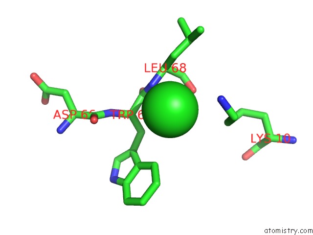
Mono view
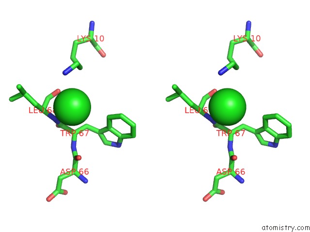
Stereo pair view
Mono view
Stereo pair view
A full contact list of Chlorine with other atoms in the Cl binding
site number 1 of A Non-Biological Atp Binding Protein with A Single Point Mutation (D65V), That Contributes to Optimized Folding and Ligand Binding within 5.0Å range:
|
Reference:
C.R.Simmons,
C.L.Magee,
D.A.Smith,
L.Lauman,
J.C.Chaput,
J.P.Allen.
Three-Dimensional Structures Reveal Multiple Adp/Atp Binding Modes For A Synthetic Class of Artificial Proteins. Biochemistry V. 49 8689 2010.
ISSN: ISSN 0006-2960
PubMed: 20822107
DOI: 10.1021/BI100398P
Page generated: Sat Jul 20 23:39:53 2024
ISSN: ISSN 0006-2960
PubMed: 20822107
DOI: 10.1021/BI100398P
Last articles
Zn in 9MJ5Zn in 9HNW
Zn in 9G0L
Zn in 9FNE
Zn in 9DZN
Zn in 9E0I
Zn in 9D32
Zn in 9DAK
Zn in 8ZXC
Zn in 8ZUF