Chlorine »
PDB 3lz1-3m98 »
3m13 »
Chlorine in PDB 3m13: Crystal Structure of the LYS265ARG Peg-Crystallized Mutant of Monomeric Sarcosine Oxidase
Enzymatic activity of Crystal Structure of the LYS265ARG Peg-Crystallized Mutant of Monomeric Sarcosine Oxidase
All present enzymatic activity of Crystal Structure of the LYS265ARG Peg-Crystallized Mutant of Monomeric Sarcosine Oxidase:
1.5.3.1;
1.5.3.1;
Protein crystallography data
The structure of Crystal Structure of the LYS265ARG Peg-Crystallized Mutant of Monomeric Sarcosine Oxidase, PDB code: 3m13
was solved by
F.S.Mathews,
Z.-W.Chen,
M.S.Jorns,
with X-Ray Crystallography technique. A brief refinement statistics is given in the table below:
| Resolution Low / High (Å) | 29.20 / 2.10 |
| Space group | P 1 21 1 |
| Cell size a, b, c (Å), α, β, γ (°) | 99.296, 69.298, 111.427, 90.00, 93.40, 90.00 |
| R / Rfree (%) | 18.7 / 24.8 |
Chlorine Binding Sites:
The binding sites of Chlorine atom in the Crystal Structure of the LYS265ARG Peg-Crystallized Mutant of Monomeric Sarcosine Oxidase
(pdb code 3m13). This binding sites where shown within
5.0 Angstroms radius around Chlorine atom.
In total 4 binding sites of Chlorine where determined in the Crystal Structure of the LYS265ARG Peg-Crystallized Mutant of Monomeric Sarcosine Oxidase, PDB code: 3m13:
Jump to Chlorine binding site number: 1; 2; 3; 4;
In total 4 binding sites of Chlorine where determined in the Crystal Structure of the LYS265ARG Peg-Crystallized Mutant of Monomeric Sarcosine Oxidase, PDB code: 3m13:
Jump to Chlorine binding site number: 1; 2; 3; 4;
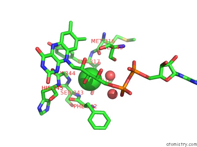
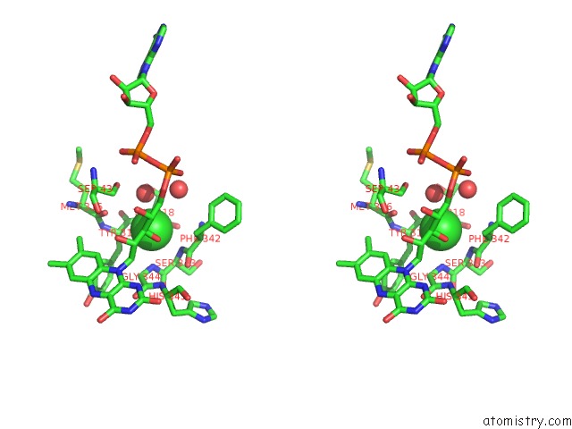
Chlorine binding site 1 out of 4 in 3m13
Go back to
Chlorine binding site 1 out
of 4 in the Crystal Structure of the LYS265ARG Peg-Crystallized Mutant of Monomeric Sarcosine Oxidase
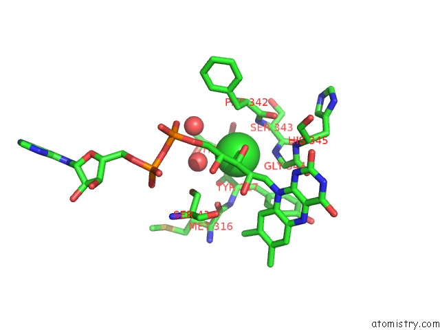
Mono view
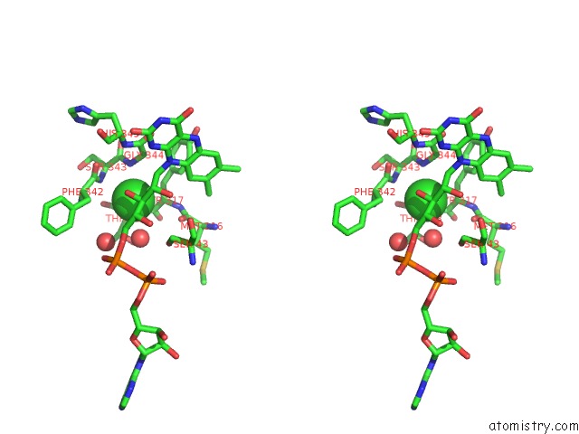
Stereo pair view
Mono view
Stereo pair view
A full contact list of Chlorine with other atoms in the Cl binding
site number 1 of Crystal Structure of the LYS265ARG Peg-Crystallized Mutant of Monomeric Sarcosine Oxidase within 5.0Å range:
|
Chlorine binding site 2 out of 4 in 3m13
Go back to
Chlorine binding site 2 out
of 4 in the Crystal Structure of the LYS265ARG Peg-Crystallized Mutant of Monomeric Sarcosine Oxidase
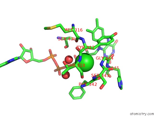
Mono view
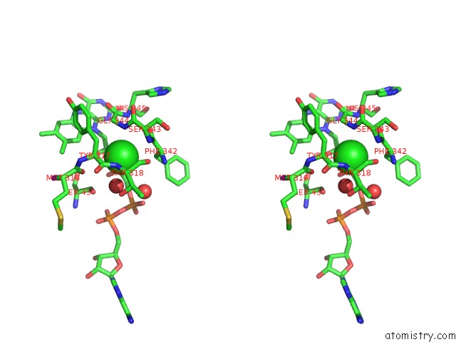
Stereo pair view
Mono view
Stereo pair view
A full contact list of Chlorine with other atoms in the Cl binding
site number 2 of Crystal Structure of the LYS265ARG Peg-Crystallized Mutant of Monomeric Sarcosine Oxidase within 5.0Å range:
|
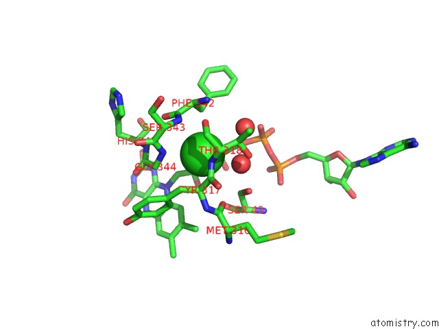
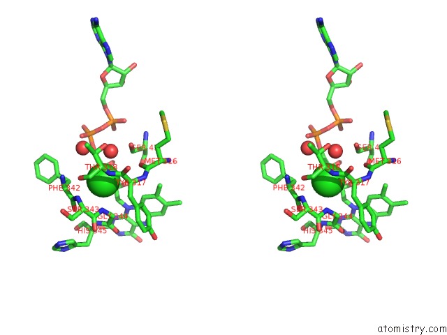
Chlorine binding site 3 out of 4 in 3m13
Go back to
Chlorine binding site 3 out
of 4 in the Crystal Structure of the LYS265ARG Peg-Crystallized Mutant of Monomeric Sarcosine Oxidase
Mono view
Stereo pair view
Mono view
Stereo pair view
A full contact list of Chlorine with other atoms in the Cl binding
site number 3 of Crystal Structure of the LYS265ARG Peg-Crystallized Mutant of Monomeric Sarcosine Oxidase within 5.0Å range:
|
Chlorine binding site 4 out of 4 in 3m13
Go back to
Chlorine binding site 4 out
of 4 in the Crystal Structure of the LYS265ARG Peg-Crystallized Mutant of Monomeric Sarcosine Oxidase
Mono view
Stereo pair view
Mono view
Stereo pair view
A full contact list of Chlorine with other atoms in the Cl binding
site number 4 of Crystal Structure of the LYS265ARG Peg-Crystallized Mutant of Monomeric Sarcosine Oxidase within 5.0Å range:
|
Reference:
M.S.Jorns,
Z.W.Chen,
F.S.Mathews.
Structural Characterization of Mutations at the Oxygen Activation Site in Monomeric Sarcosine Oxidase. Biochemistry V. 49 3631 2010.
ISSN: ISSN 0006-2960
PubMed: 20353187
DOI: 10.1021/BI100160J
Page generated: Sat Jul 20 23:54:06 2024
ISSN: ISSN 0006-2960
PubMed: 20353187
DOI: 10.1021/BI100160J
Last articles
Zn in 9J0NZn in 9J0O
Zn in 9J0P
Zn in 9FJX
Zn in 9EKB
Zn in 9C0F
Zn in 9CAH
Zn in 9CH0
Zn in 9CH3
Zn in 9CH1