Chlorine »
PDB 3lz1-3m98 »
3m5v »
Chlorine in PDB 3m5v: Crystal Structure of Dihydrodipicolinate Synthase From Campylobacter Jejuni
Enzymatic activity of Crystal Structure of Dihydrodipicolinate Synthase From Campylobacter Jejuni
All present enzymatic activity of Crystal Structure of Dihydrodipicolinate Synthase From Campylobacter Jejuni:
4.2.1.52;
4.2.1.52;
Protein crystallography data
The structure of Crystal Structure of Dihydrodipicolinate Synthase From Campylobacter Jejuni, PDB code: 3m5v
was solved by
Y.Kim,
M.Zhou,
K.Kwon,
W.F.Anderson,
A.Joachimiak,
Center For Structuralgenomics Of Infectious Diseases (Csgid),
with X-Ray Crystallography technique. A brief refinement statistics is given in the table below:
| Resolution Low / High (Å) | 43.06 / 1.80 |
| Space group | P 21 21 21 |
| Cell size a, b, c (Å), α, β, γ (°) | 72.360, 86.032, 198.948, 90.00, 90.00, 90.00 |
| R / Rfree (%) | 14.6 / 18.1 |
Chlorine Binding Sites:
The binding sites of Chlorine atom in the Crystal Structure of Dihydrodipicolinate Synthase From Campylobacter Jejuni
(pdb code 3m5v). This binding sites where shown within
5.0 Angstroms radius around Chlorine atom.
In total 10 binding sites of Chlorine where determined in the Crystal Structure of Dihydrodipicolinate Synthase From Campylobacter Jejuni, PDB code: 3m5v:
Jump to Chlorine binding site number: 1; 2; 3; 4; 5; 6; 7; 8; 9; 10;
In total 10 binding sites of Chlorine where determined in the Crystal Structure of Dihydrodipicolinate Synthase From Campylobacter Jejuni, PDB code: 3m5v:
Jump to Chlorine binding site number: 1; 2; 3; 4; 5; 6; 7; 8; 9; 10;
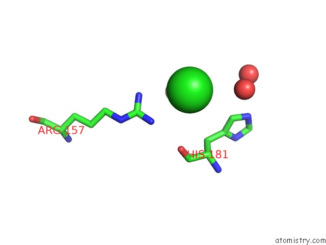
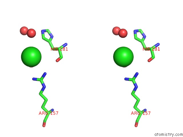
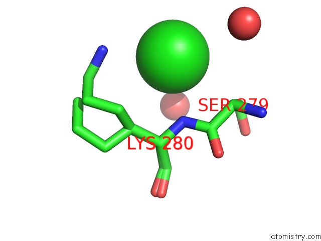
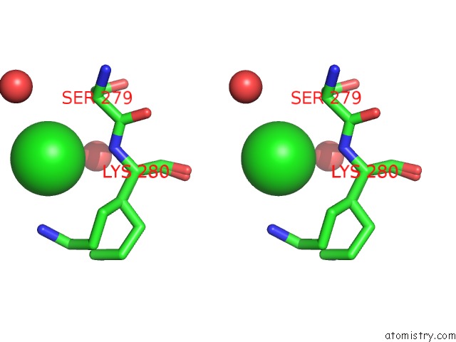
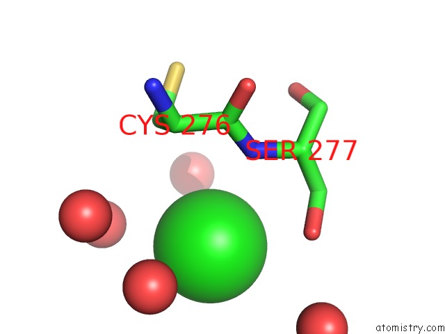
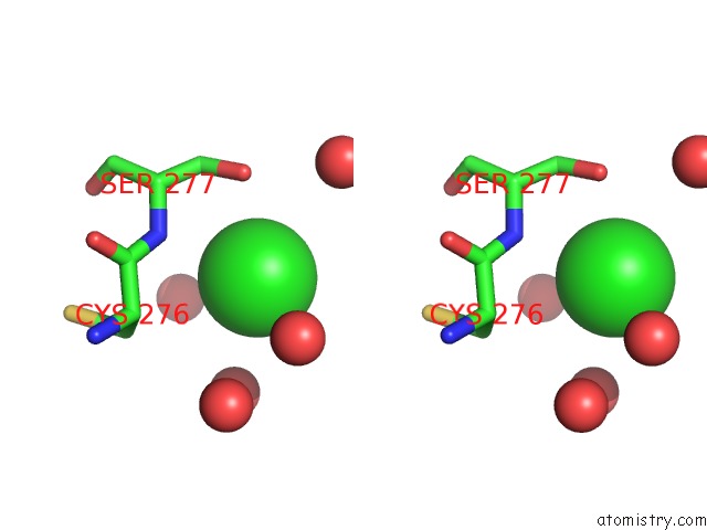
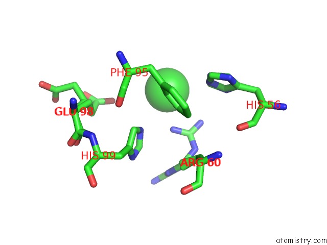
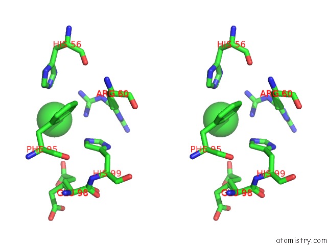
Chlorine binding site 1 out of 10 in 3m5v
Go back to
Chlorine binding site 1 out
of 10 in the Crystal Structure of Dihydrodipicolinate Synthase From Campylobacter Jejuni
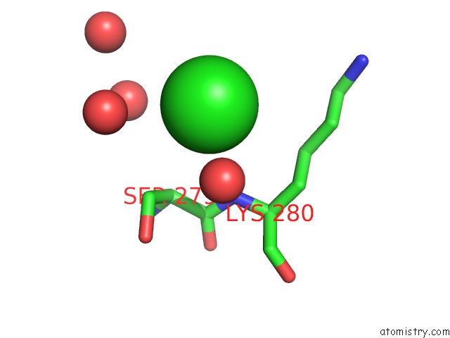
Mono view
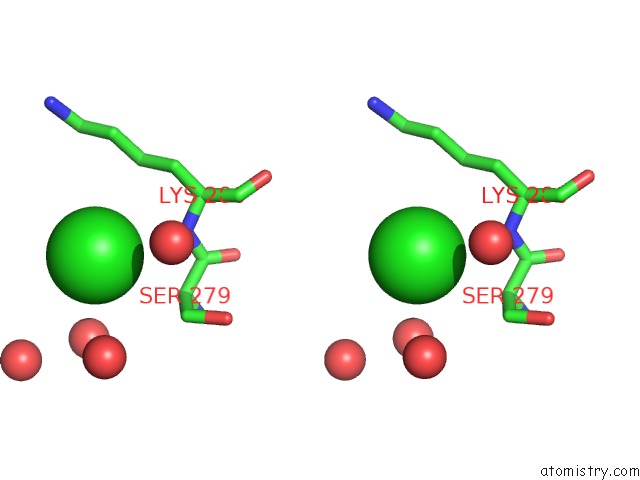
Stereo pair view
Mono view
Stereo pair view
A full contact list of Chlorine with other atoms in the Cl binding
site number 1 of Crystal Structure of Dihydrodipicolinate Synthase From Campylobacter Jejuni within 5.0Å range:
|
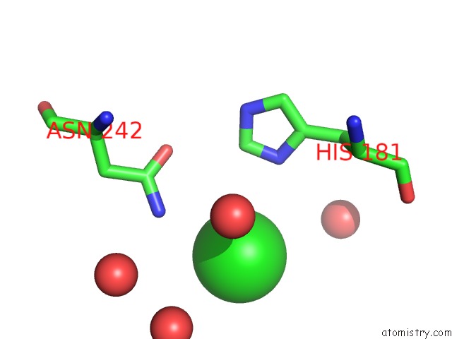
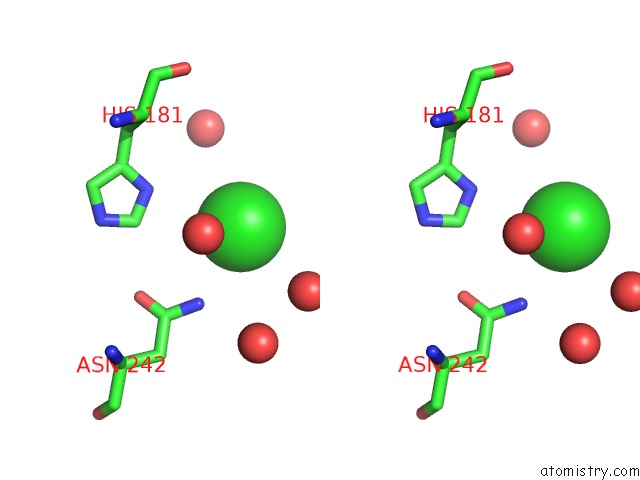
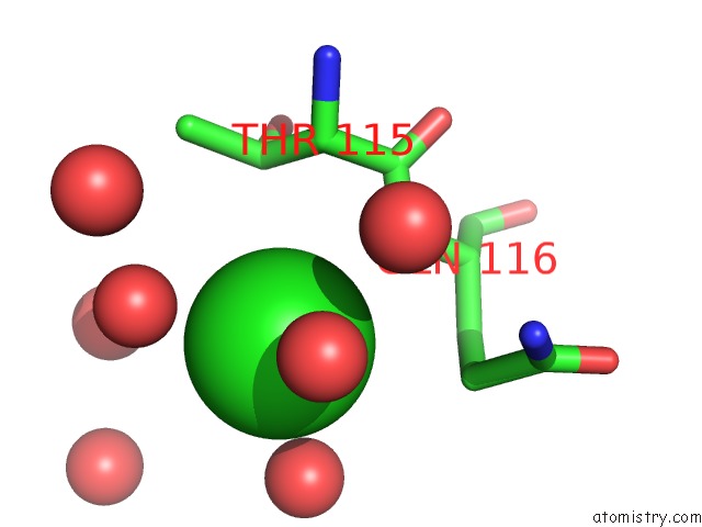
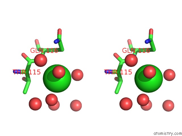
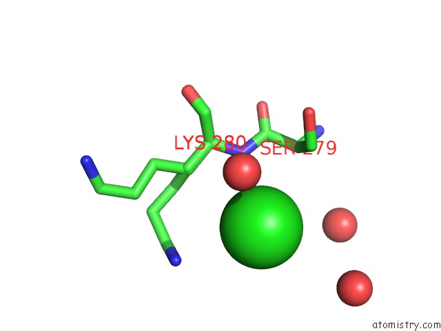
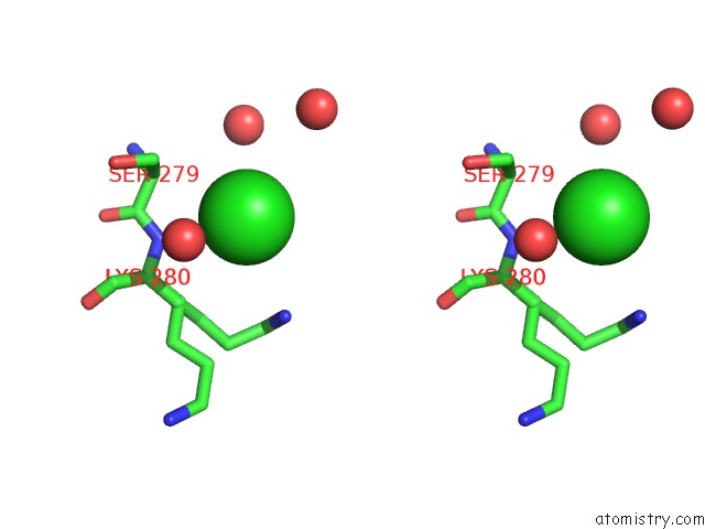
Chlorine binding site 2 out of 10 in 3m5v
Go back to
Chlorine binding site 2 out
of 10 in the Crystal Structure of Dihydrodipicolinate Synthase From Campylobacter Jejuni
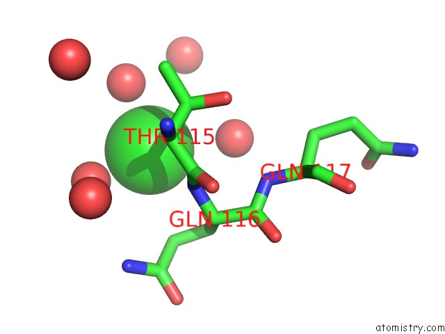
Mono view
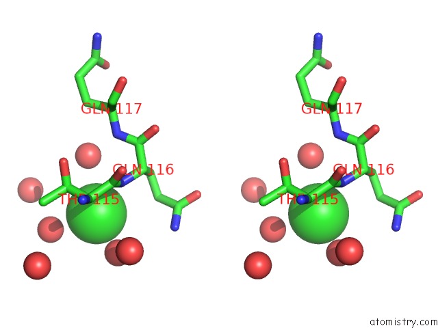
Stereo pair view
Mono view
Stereo pair view
A full contact list of Chlorine with other atoms in the Cl binding
site number 2 of Crystal Structure of Dihydrodipicolinate Synthase From Campylobacter Jejuni within 5.0Å range:
|
Chlorine binding site 3 out of 10 in 3m5v
Go back to
Chlorine binding site 3 out
of 10 in the Crystal Structure of Dihydrodipicolinate Synthase From Campylobacter Jejuni
Mono view
Stereo pair view
Mono view
Stereo pair view
A full contact list of Chlorine with other atoms in the Cl binding
site number 3 of Crystal Structure of Dihydrodipicolinate Synthase From Campylobacter Jejuni within 5.0Å range:
|
Chlorine binding site 4 out of 10 in 3m5v
Go back to
Chlorine binding site 4 out
of 10 in the Crystal Structure of Dihydrodipicolinate Synthase From Campylobacter Jejuni
Mono view
Stereo pair view
Mono view
Stereo pair view
A full contact list of Chlorine with other atoms in the Cl binding
site number 4 of Crystal Structure of Dihydrodipicolinate Synthase From Campylobacter Jejuni within 5.0Å range:
|
Chlorine binding site 5 out of 10 in 3m5v
Go back to
Chlorine binding site 5 out
of 10 in the Crystal Structure of Dihydrodipicolinate Synthase From Campylobacter Jejuni
Mono view
Stereo pair view
Mono view
Stereo pair view
A full contact list of Chlorine with other atoms in the Cl binding
site number 5 of Crystal Structure of Dihydrodipicolinate Synthase From Campylobacter Jejuni within 5.0Å range:
|
Chlorine binding site 6 out of 10 in 3m5v
Go back to
Chlorine binding site 6 out
of 10 in the Crystal Structure of Dihydrodipicolinate Synthase From Campylobacter Jejuni
Mono view
Stereo pair view
Mono view
Stereo pair view
A full contact list of Chlorine with other atoms in the Cl binding
site number 6 of Crystal Structure of Dihydrodipicolinate Synthase From Campylobacter Jejuni within 5.0Å range:
|
Chlorine binding site 7 out of 10 in 3m5v
Go back to
Chlorine binding site 7 out
of 10 in the Crystal Structure of Dihydrodipicolinate Synthase From Campylobacter Jejuni
Mono view
Stereo pair view
Mono view
Stereo pair view
A full contact list of Chlorine with other atoms in the Cl binding
site number 7 of Crystal Structure of Dihydrodipicolinate Synthase From Campylobacter Jejuni within 5.0Å range:
|
Chlorine binding site 8 out of 10 in 3m5v
Go back to
Chlorine binding site 8 out
of 10 in the Crystal Structure of Dihydrodipicolinate Synthase From Campylobacter Jejuni
Mono view
Stereo pair view
Mono view
Stereo pair view
A full contact list of Chlorine with other atoms in the Cl binding
site number 8 of Crystal Structure of Dihydrodipicolinate Synthase From Campylobacter Jejuni within 5.0Å range:
|
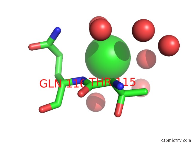
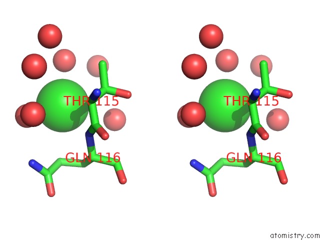
Chlorine binding site 9 out of 10 in 3m5v
Go back to
Chlorine binding site 9 out
of 10 in the Crystal Structure of Dihydrodipicolinate Synthase From Campylobacter Jejuni
Mono view
Stereo pair view
Mono view
Stereo pair view
A full contact list of Chlorine with other atoms in the Cl binding
site number 9 of Crystal Structure of Dihydrodipicolinate Synthase From Campylobacter Jejuni within 5.0Å range:
|
Chlorine binding site 10 out of 10 in 3m5v
Go back to
Chlorine binding site 10 out
of 10 in the Crystal Structure of Dihydrodipicolinate Synthase From Campylobacter Jejuni
Mono view
Stereo pair view
Mono view
Stereo pair view
A full contact list of Chlorine with other atoms in the Cl binding
site number 10 of Crystal Structure of Dihydrodipicolinate Synthase From Campylobacter Jejuni within 5.0Å range:
|
Reference:
Y.Kim,
M.Zhou,
K.Kwon,
W.F.Anderson,
A.Joachimiak,
Center For Structural Genomics Of Infectious Diseases(Csgid).
Crystal Structure of Dihydrodipicolinate Synthase From Campylobacter Jejuni To Be Published.
Page generated: Sun Jul 21 00:00:00 2024
Last articles
Zn in 9MJ5Zn in 9HNW
Zn in 9G0L
Zn in 9FNE
Zn in 9DZN
Zn in 9E0I
Zn in 9D32
Zn in 9DAK
Zn in 8ZXC
Zn in 8ZUF