Chlorine »
PDB 3lz1-3m98 »
3m6z »
Chlorine in PDB 3m6z: Crystal Structure of An N-Terminal 44 kDa Fragment of Topoisomerase V in the Presence of Guanidium Hydrochloride
Protein crystallography data
The structure of Crystal Structure of An N-Terminal 44 kDa Fragment of Topoisomerase V in the Presence of Guanidium Hydrochloride, PDB code: 3m6z
was solved by
R.Rajan,
B.Taneja,
A.Mondragon,
with X-Ray Crystallography technique. A brief refinement statistics is given in the table below:
| Resolution Low / High (Å) | 28.90 / 1.40 |
| Space group | P 21 21 21 |
| Cell size a, b, c (Å), α, β, γ (°) | 63.640, 80.110, 137.220, 90.00, 90.00, 90.00 |
| R / Rfree (%) | 16.5 / 18.4 |
Other elements in 3m6z:
The structure of Crystal Structure of An N-Terminal 44 kDa Fragment of Topoisomerase V in the Presence of Guanidium Hydrochloride also contains other interesting chemical elements:
| Magnesium | (Mg) | 3 atoms |
Chlorine Binding Sites:
The binding sites of Chlorine atom in the Crystal Structure of An N-Terminal 44 kDa Fragment of Topoisomerase V in the Presence of Guanidium Hydrochloride
(pdb code 3m6z). This binding sites where shown within
5.0 Angstroms radius around Chlorine atom.
In total 2 binding sites of Chlorine where determined in the Crystal Structure of An N-Terminal 44 kDa Fragment of Topoisomerase V in the Presence of Guanidium Hydrochloride, PDB code: 3m6z:
Jump to Chlorine binding site number: 1; 2;
In total 2 binding sites of Chlorine where determined in the Crystal Structure of An N-Terminal 44 kDa Fragment of Topoisomerase V in the Presence of Guanidium Hydrochloride, PDB code: 3m6z:
Jump to Chlorine binding site number: 1; 2;
Chlorine binding site 1 out of 2 in 3m6z
Go back to
Chlorine binding site 1 out
of 2 in the Crystal Structure of An N-Terminal 44 kDa Fragment of Topoisomerase V in the Presence of Guanidium Hydrochloride
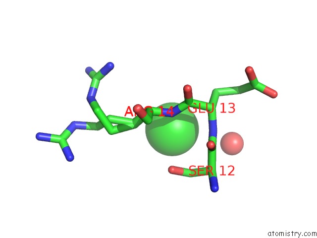
Mono view
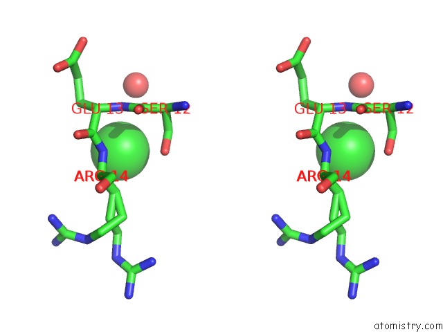
Stereo pair view
Mono view
Stereo pair view
A full contact list of Chlorine with other atoms in the Cl binding
site number 1 of Crystal Structure of An N-Terminal 44 kDa Fragment of Topoisomerase V in the Presence of Guanidium Hydrochloride within 5.0Å range:
|
Chlorine binding site 2 out of 2 in 3m6z
Go back to
Chlorine binding site 2 out
of 2 in the Crystal Structure of An N-Terminal 44 kDa Fragment of Topoisomerase V in the Presence of Guanidium Hydrochloride
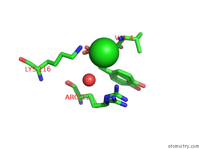
Mono view
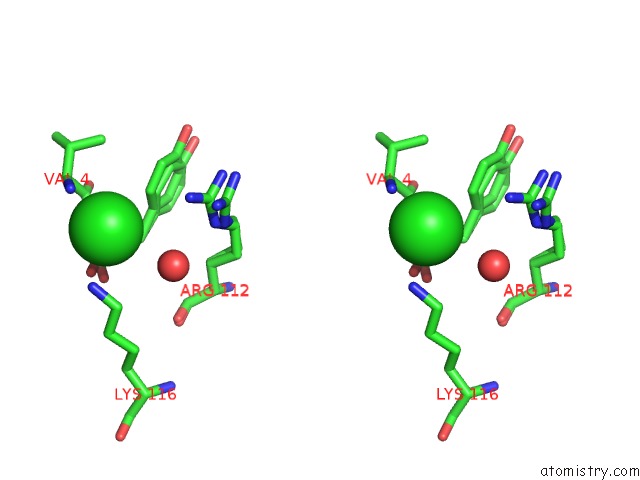
Stereo pair view
Mono view
Stereo pair view
A full contact list of Chlorine with other atoms in the Cl binding
site number 2 of Crystal Structure of An N-Terminal 44 kDa Fragment of Topoisomerase V in the Presence of Guanidium Hydrochloride within 5.0Å range:
|
Reference:
R.Rajan,
B.Taneja,
A.Mondragon.
Structures of Minimal Catalytic Fragments of Topoisomerase V Reveals Conformational Changes Relevant For Dna Binding. Structure V. 18 829 2010.
ISSN: ISSN 0969-2126
PubMed: 20637419
DOI: 10.1016/J.STR.2010.03.006
Page generated: Fri Jul 11 07:47:01 2025
ISSN: ISSN 0969-2126
PubMed: 20637419
DOI: 10.1016/J.STR.2010.03.006
Last articles
Fe in 2YXOFe in 2YRS
Fe in 2YXC
Fe in 2YNM
Fe in 2YVJ
Fe in 2YP1
Fe in 2YU2
Fe in 2YU1
Fe in 2YQB
Fe in 2YOO