Chlorine »
PDB 3m9f-3mle »
3mc5 »
Chlorine in PDB 3mc5: Human Aldose Reductase Mutant T113V in Complex with IDD393 Crystallized in Spacegroup P1
Enzymatic activity of Human Aldose Reductase Mutant T113V in Complex with IDD393 Crystallized in Spacegroup P1
All present enzymatic activity of Human Aldose Reductase Mutant T113V in Complex with IDD393 Crystallized in Spacegroup P1:
1.1.1.21;
1.1.1.21;
Protein crystallography data
The structure of Human Aldose Reductase Mutant T113V in Complex with IDD393 Crystallized in Spacegroup P1, PDB code: 3mc5
was solved by
C.Koch,
A.Heine,
G.Klebe,
with X-Ray Crystallography technique. A brief refinement statistics is given in the table below:
| Resolution Low / High (Å) | 10.00 / 1.14 |
| Space group | P 1 |
| Cell size a, b, c (Å), α, β, γ (°) | 40.150, 47.190, 47.300, 76.10, 67.54, 76.97 |
| R / Rfree (%) | 12.3 / 15.8 |
Chlorine Binding Sites:
The binding sites of Chlorine atom in the Human Aldose Reductase Mutant T113V in Complex with IDD393 Crystallized in Spacegroup P1
(pdb code 3mc5). This binding sites where shown within
5.0 Angstroms radius around Chlorine atom.
In total only one binding site of Chlorine was determined in the Human Aldose Reductase Mutant T113V in Complex with IDD393 Crystallized in Spacegroup P1, PDB code: 3mc5:
In total only one binding site of Chlorine was determined in the Human Aldose Reductase Mutant T113V in Complex with IDD393 Crystallized in Spacegroup P1, PDB code: 3mc5:
Chlorine binding site 1 out of 1 in 3mc5
Go back to
Chlorine binding site 1 out
of 1 in the Human Aldose Reductase Mutant T113V in Complex with IDD393 Crystallized in Spacegroup P1
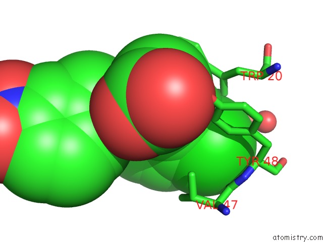
Mono view
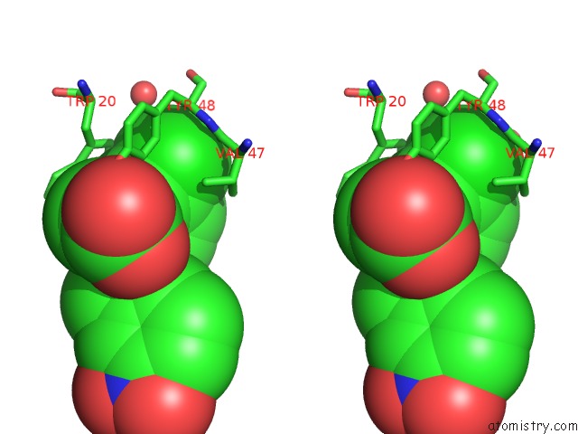
Stereo pair view
Mono view
Stereo pair view
A full contact list of Chlorine with other atoms in the Cl binding
site number 1 of Human Aldose Reductase Mutant T113V in Complex with IDD393 Crystallized in Spacegroup P1 within 5.0Å range:
|
Reference:
C.Koch,
A.Heine,
G.Klebe.
Ligand-Induced Fit Affects Binding Modes and Provokes Changes in Crystal Packing of Aldose Reductase Biochim.Biophys.Acta V.1810 879 2011.
ISSN: ISSN 0006-3002
PubMed: 21684320
DOI: 10.1016/J.BBAGEN.2011.06.001
Page generated: Sun Jul 21 00:07:30 2024
ISSN: ISSN 0006-3002
PubMed: 21684320
DOI: 10.1016/J.BBAGEN.2011.06.001
Last articles
Zn in 9J0NZn in 9J0O
Zn in 9J0P
Zn in 9FJX
Zn in 9EKB
Zn in 9C0F
Zn in 9CAH
Zn in 9CH0
Zn in 9CH3
Zn in 9CH1