Chlorine »
PDB 3n4e-3nb5 »
3n85 »
Chlorine in PDB 3n85: Crystallographic Trimer of HER2 Extracellular Regions in Complex with Tryptophan-Rich Antibody Fragment
Enzymatic activity of Crystallographic Trimer of HER2 Extracellular Regions in Complex with Tryptophan-Rich Antibody Fragment
All present enzymatic activity of Crystallographic Trimer of HER2 Extracellular Regions in Complex with Tryptophan-Rich Antibody Fragment:
2.7.10.1;
2.7.10.1;
Protein crystallography data
The structure of Crystallographic Trimer of HER2 Extracellular Regions in Complex with Tryptophan-Rich Antibody Fragment, PDB code: 3n85
was solved by
C.Eigenbrot,
with X-Ray Crystallography technique. A brief refinement statistics is given in the table below:
| Resolution Low / High (Å) | 50.00 / 3.20 |
| Space group | P 63 2 2 |
| Cell size a, b, c (Å), α, β, γ (°) | 182.220, 182.220, 330.851, 90.00, 90.00, 120.00 |
| R / Rfree (%) | 20.6 / 23 |
Chlorine Binding Sites:
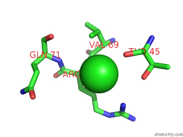
The binding sites of Chlorine atom in the Crystallographic Trimer of HER2 Extracellular Regions in Complex with Tryptophan-Rich Antibody Fragment
(pdb code 3n85). This binding sites where shown within
5.0 Angstroms radius around Chlorine atom.
In total only one binding site of Chlorine was determined in the Crystallographic Trimer of HER2 Extracellular Regions in Complex with Tryptophan-Rich Antibody Fragment, PDB code: 3n85:
In total only one binding site of Chlorine was determined in the Crystallographic Trimer of HER2 Extracellular Regions in Complex with Tryptophan-Rich Antibody Fragment, PDB code: 3n85:
Chlorine binding site 1 out of 1 in 3n85
Go back to
Chlorine binding site 1 out
of 1 in the Crystallographic Trimer of HER2 Extracellular Regions in Complex with Tryptophan-Rich Antibody Fragment
Mono view
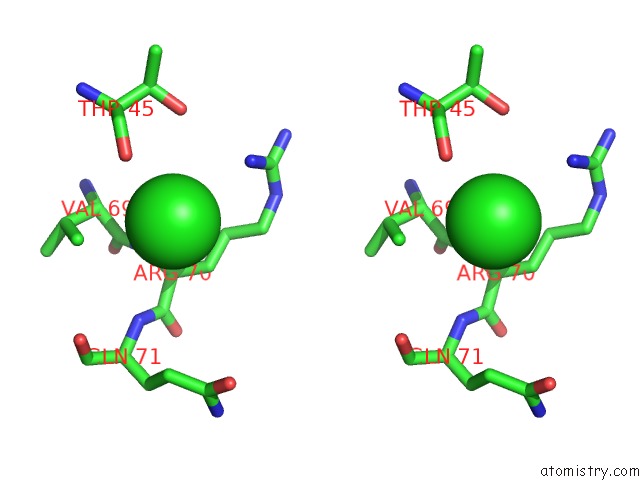
Stereo pair view
Mono view
Stereo pair view
A full contact list of Chlorine with other atoms in the Cl binding
site number 1 of Crystallographic Trimer of HER2 Extracellular Regions in Complex with Tryptophan-Rich Antibody Fragment within 5.0Å range:
|
Reference:
R.D.Fisher,
M.Ultsch,
A.Lingel,
G.Schaefer,
L.Shao,
S.Birtalan,
S.S.Sidhu,
C.Eigenbrot.
Structure of the Complex Between HER2 and An Antibody Paratope Formed By Side Chains From Tryptophan and Serine. J.Mol.Biol. V. 402 217 2010.
ISSN: ISSN 0022-2836
PubMed: 20654626
DOI: 10.1016/J.JMB.2010.07.027
Page generated: Sun Jul 21 00:42:14 2024
ISSN: ISSN 0022-2836
PubMed: 20654626
DOI: 10.1016/J.JMB.2010.07.027
Last articles
Zn in 9J0NZn in 9J0O
Zn in 9J0P
Zn in 9FJX
Zn in 9EKB
Zn in 9C0F
Zn in 9CAH
Zn in 9CH0
Zn in 9CH3
Zn in 9CH1