Chlorine »
PDB 3nbu-3nnw »
3nbu »
Chlorine in PDB 3nbu: Crystal Structure of Pgi Glucosephosphate Isomerase
Enzymatic activity of Crystal Structure of Pgi Glucosephosphate Isomerase
All present enzymatic activity of Crystal Structure of Pgi Glucosephosphate Isomerase:
5.3.1.9;
5.3.1.9;
Protein crystallography data
The structure of Crystal Structure of Pgi Glucosephosphate Isomerase, PDB code: 3nbu
was solved by
T.Alber,
C.Zubieta,
M.Totir,
A.May,
N.Echols,
with X-Ray Crystallography technique. A brief refinement statistics is given in the table below:
| Resolution Low / High (Å) | 89.00 / 2.05 |
| Space group | P 1 |
| Cell size a, b, c (Å), α, β, γ (°) | 69.806, 72.874, 181.851, 92.47, 97.82, 114.57 |
| R / Rfree (%) | 16.9 / 23 |
Chlorine Binding Sites:
The binding sites of Chlorine atom in the Crystal Structure of Pgi Glucosephosphate Isomerase
(pdb code 3nbu). This binding sites where shown within
5.0 Angstroms radius around Chlorine atom.
In total only one binding site of Chlorine was determined in the Crystal Structure of Pgi Glucosephosphate Isomerase, PDB code: 3nbu:
In total only one binding site of Chlorine was determined in the Crystal Structure of Pgi Glucosephosphate Isomerase, PDB code: 3nbu:
Chlorine binding site 1 out of 1 in 3nbu
Go back to
Chlorine binding site 1 out
of 1 in the Crystal Structure of Pgi Glucosephosphate Isomerase
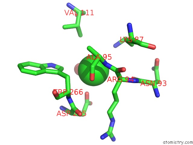
Mono view
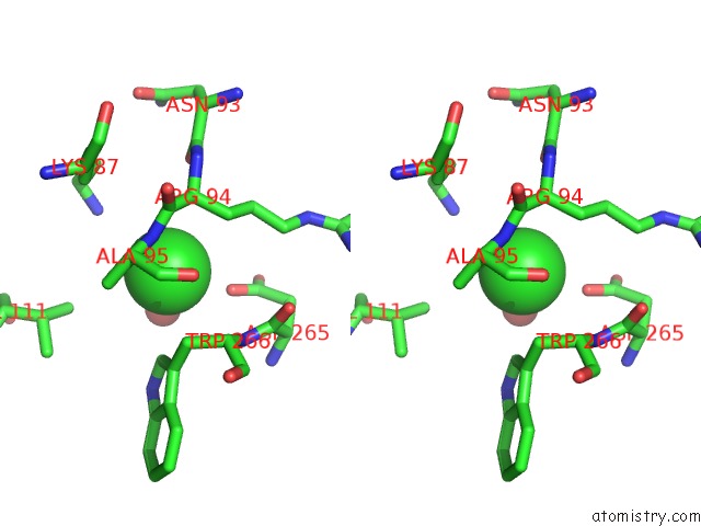
Stereo pair view
Mono view
Stereo pair view
A full contact list of Chlorine with other atoms in the Cl binding
site number 1 of Crystal Structure of Pgi Glucosephosphate Isomerase within 5.0Å range:
|
Reference:
M.Totir,
N.Echols,
M.Nanao,
C.L.Gee,
A.Moskaleva,
S.Gradia,
A.T.Iavarone,
J.M.Berger,
A.P.May,
C.Zubieta,
T.Alber.
Macro-to-Micro Structural Proteomics: Native Source Proteins For High-Throughput Crystallization. Plos One V. 7 32498 2012.
ISSN: ESSN 1932-6203
PubMed: 22393408
DOI: 10.1371/JOURNAL.PONE.0032498
Page generated: Sun Jul 21 00:56:50 2024
ISSN: ESSN 1932-6203
PubMed: 22393408
DOI: 10.1371/JOURNAL.PONE.0032498
Last articles
Zn in 9J0NZn in 9J0O
Zn in 9J0P
Zn in 9FJX
Zn in 9EKB
Zn in 9C0F
Zn in 9CAH
Zn in 9CH0
Zn in 9CH3
Zn in 9CH1