Chlorine »
PDB 3nnw-3nvu »
3nsj »
Chlorine in PDB 3nsj: The X-Ray Crystal Structure of Lymphocyte Perforin
Protein crystallography data
The structure of The X-Ray Crystal Structure of Lymphocyte Perforin, PDB code: 3nsj
was solved by
R.H.Law,
J.C.Whisstock,
T.T.Caradoc-Davies,
with X-Ray Crystallography technique. A brief refinement statistics is given in the table below:
| Resolution Low / High (Å) | 47.36 / 2.75 |
| Space group | P 21 21 21 |
| Cell size a, b, c (Å), α, β, γ (°) | 78.640, 109.800, 141.030, 90.00, 90.00, 90.00 |
| R / Rfree (%) | 19.6 / 21.5 |
Other elements in 3nsj:
The structure of The X-Ray Crystal Structure of Lymphocyte Perforin also contains other interesting chemical elements:
| Iodine | (I) | 4 atoms |
| Calcium | (Ca) | 2 atoms |
Chlorine Binding Sites:
The binding sites of Chlorine atom in the The X-Ray Crystal Structure of Lymphocyte Perforin
(pdb code 3nsj). This binding sites where shown within
5.0 Angstroms radius around Chlorine atom.
In total 2 binding sites of Chlorine where determined in the The X-Ray Crystal Structure of Lymphocyte Perforin, PDB code: 3nsj:
Jump to Chlorine binding site number: 1; 2;
In total 2 binding sites of Chlorine where determined in the The X-Ray Crystal Structure of Lymphocyte Perforin, PDB code: 3nsj:
Jump to Chlorine binding site number: 1; 2;
Chlorine binding site 1 out of 2 in 3nsj
Go back to
Chlorine binding site 1 out
of 2 in the The X-Ray Crystal Structure of Lymphocyte Perforin
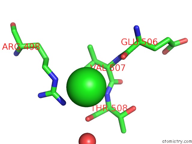
Mono view
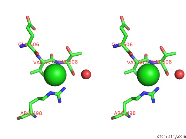
Stereo pair view
Mono view
Stereo pair view
A full contact list of Chlorine with other atoms in the Cl binding
site number 1 of The X-Ray Crystal Structure of Lymphocyte Perforin within 5.0Å range:
|
Chlorine binding site 2 out of 2 in 3nsj
Go back to
Chlorine binding site 2 out
of 2 in the The X-Ray Crystal Structure of Lymphocyte Perforin
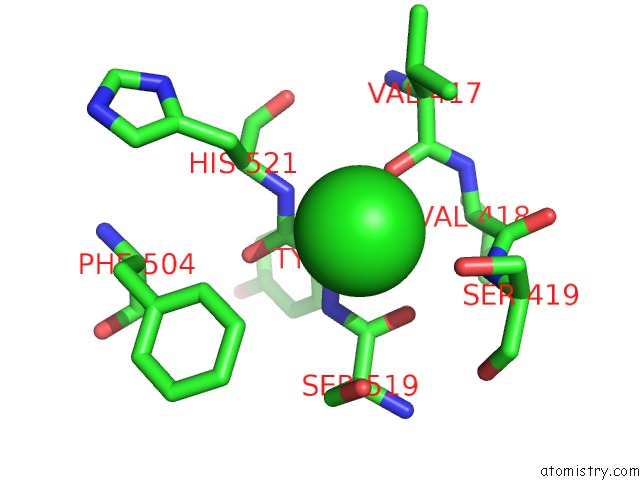
Mono view
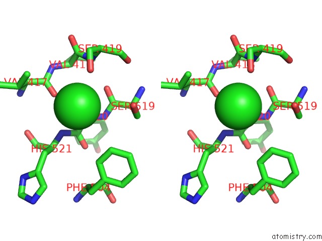
Stereo pair view
Mono view
Stereo pair view
A full contact list of Chlorine with other atoms in the Cl binding
site number 2 of The X-Ray Crystal Structure of Lymphocyte Perforin within 5.0Å range:
|
Reference:
R.H.Law,
N.Lukoyanova,
I.Voskoboinik,
T.T.Caradoc-Davies,
K.Baran,
M.A.Dunstone,
M.E.D'angelo,
E.V.Orlova,
F.Coulibaly,
S.Verschoor,
K.A.Browne,
A.Ciccone,
M.J.Kuiper,
P.I.Bird,
J.A.Trapani,
H.R.Saibil,
J.C.Whisstock.
The Structural Basis For Membrane Binding and Pore Formation By Lymphocyte Perforin. Nature V. 468 447 2010.
ISSN: ISSN 0028-0836
PubMed: 21037563
DOI: 10.1038/NATURE09518
Page generated: Sun Jul 21 01:12:17 2024
ISSN: ISSN 0028-0836
PubMed: 21037563
DOI: 10.1038/NATURE09518
Last articles
Zn in 9JYWZn in 9IR4
Zn in 9IR3
Zn in 9GMX
Zn in 9GMW
Zn in 9JEJ
Zn in 9ERF
Zn in 9ERE
Zn in 9EGV
Zn in 9EGW