Chlorine »
PDB 3nw9-3o6j »
3o4d »
Chlorine in PDB 3o4d: Crystal Structure of Symfoil-4P: De Novo Designed Beta-Trefoil Architecture with Symmetric Primary Structure
Protein crystallography data
The structure of Crystal Structure of Symfoil-4P: De Novo Designed Beta-Trefoil Architecture with Symmetric Primary Structure, PDB code: 3o4d
was solved by
J.Lee,
M.Blaber,
with X-Ray Crystallography technique. A brief refinement statistics is given in the table below:
| Resolution Low / High (Å) | 42.39 / 1.65 |
| Space group | I 2 2 2 |
| Cell size a, b, c (Å), α, β, γ (°) | 50.372, 53.151, 84.780, 90.00, 90.00, 90.00 |
| R / Rfree (%) | 19 / 22.6 |
Chlorine Binding Sites:
The binding sites of Chlorine atom in the Crystal Structure of Symfoil-4P: De Novo Designed Beta-Trefoil Architecture with Symmetric Primary Structure
(pdb code 3o4d). This binding sites where shown within
5.0 Angstroms radius around Chlorine atom.
In total only one binding site of Chlorine was determined in the Crystal Structure of Symfoil-4P: De Novo Designed Beta-Trefoil Architecture with Symmetric Primary Structure, PDB code: 3o4d:
In total only one binding site of Chlorine was determined in the Crystal Structure of Symfoil-4P: De Novo Designed Beta-Trefoil Architecture with Symmetric Primary Structure, PDB code: 3o4d:
Chlorine binding site 1 out of 1 in 3o4d
Go back to
Chlorine binding site 1 out
of 1 in the Crystal Structure of Symfoil-4P: De Novo Designed Beta-Trefoil Architecture with Symmetric Primary Structure
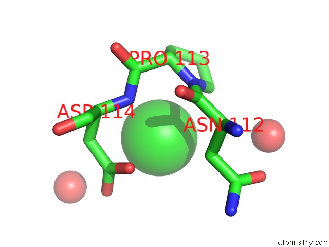
Mono view
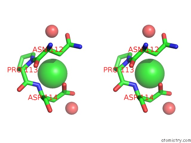
Stereo pair view
Mono view
Stereo pair view
A full contact list of Chlorine with other atoms in the Cl binding
site number 1 of Crystal Structure of Symfoil-4P: De Novo Designed Beta-Trefoil Architecture with Symmetric Primary Structure within 5.0Å range:
|
Reference:
J.Lee,
M.Blaber.
Experimental Support For the Evolution of Symmetric Protein Architecture From A Simple Peptide Motif. Proc.Natl.Acad.Sci.Usa V. 108 126 2011.
ISSN: ISSN 0027-8424
PubMed: 21173271
DOI: 10.1073/PNAS.1015032108
Page generated: Sun Jul 21 01:23:54 2024
ISSN: ISSN 0027-8424
PubMed: 21173271
DOI: 10.1073/PNAS.1015032108
Last articles
Zn in 9J0NZn in 9J0O
Zn in 9J0P
Zn in 9FJX
Zn in 9EKB
Zn in 9C0F
Zn in 9CAH
Zn in 9CH0
Zn in 9CH3
Zn in 9CH1