Chlorine »
PDB 3oid-3oow »
3oin »
Chlorine in PDB 3oin: Crystal Structure of Saccharomyces Cerevisiae NEP1/EMG1 Bound to S- Adenosylhomocysteine and 1 Molecule of Cognate Rna
Protein crystallography data
The structure of Crystal Structure of Saccharomyces Cerevisiae NEP1/EMG1 Bound to S- Adenosylhomocysteine and 1 Molecule of Cognate Rna, PDB code: 3oin
was solved by
S.R.Thomas,
N.Laronde-Leblanc,
with X-Ray Crystallography technique. A brief refinement statistics is given in the table below:
| Resolution Low / High (Å) | 27.68 / 1.90 |
| Space group | P 21 21 21 |
| Cell size a, b, c (Å), α, β, γ (°) | 44.774, 88.749, 115.751, 90.00, 90.00, 90.00 |
| R / Rfree (%) | 19.2 / 23 |
Other elements in 3oin:
The structure of Crystal Structure of Saccharomyces Cerevisiae NEP1/EMG1 Bound to S- Adenosylhomocysteine and 1 Molecule of Cognate Rna also contains other interesting chemical elements:
| Magnesium | (Mg) | 1 atom |
Chlorine Binding Sites:
The binding sites of Chlorine atom in the Crystal Structure of Saccharomyces Cerevisiae NEP1/EMG1 Bound to S- Adenosylhomocysteine and 1 Molecule of Cognate Rna
(pdb code 3oin). This binding sites where shown within
5.0 Angstroms radius around Chlorine atom.
In total only one binding site of Chlorine was determined in the Crystal Structure of Saccharomyces Cerevisiae NEP1/EMG1 Bound to S- Adenosylhomocysteine and 1 Molecule of Cognate Rna, PDB code: 3oin:
In total only one binding site of Chlorine was determined in the Crystal Structure of Saccharomyces Cerevisiae NEP1/EMG1 Bound to S- Adenosylhomocysteine and 1 Molecule of Cognate Rna, PDB code: 3oin:
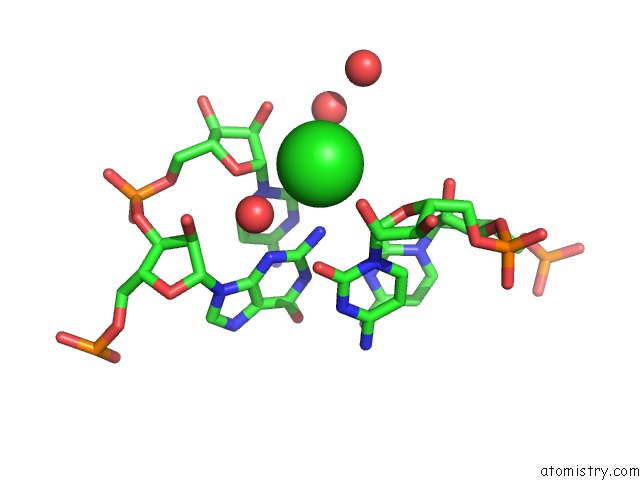
Chlorine binding site 1 out of 1 in 3oin
Go back to
Chlorine binding site 1 out
of 1 in the Crystal Structure of Saccharomyces Cerevisiae NEP1/EMG1 Bound to S- Adenosylhomocysteine and 1 Molecule of Cognate Rna
Mono view

Stereo pair view
Mono view
Stereo pair view
A full contact list of Chlorine with other atoms in the Cl binding
site number 1 of Crystal Structure of Saccharomyces Cerevisiae NEP1/EMG1 Bound to S- Adenosylhomocysteine and 1 Molecule of Cognate Rna within 5.0Å range:
|
Reference:
S.R.Thomas,
C.A.Keller,
A.Szyk,
J.R.Cannon,
N.A.Laronde-Leblanc.
Structural Insight Into the Functional Mechanism of NEP1/EMG1 N1-Specific Pseudouridine Methyltransferase in Ribosome Biogenesis. Nucleic Acids Res. V. 39 2445 2011.
ISSN: ISSN 0305-1048
PubMed: 21087996
DOI: 10.1093/NAR/GKQ1131
Page generated: Sun Jul 21 01:39:54 2024
ISSN: ISSN 0305-1048
PubMed: 21087996
DOI: 10.1093/NAR/GKQ1131
Last articles
Zn in 9J0NZn in 9J0O
Zn in 9J0P
Zn in 9FJX
Zn in 9EKB
Zn in 9C0F
Zn in 9CAH
Zn in 9CH0
Zn in 9CH3
Zn in 9CH1