Chlorine »
PDB 3oid-3oow »
3omu »
Chlorine in PDB 3omu: Crystal Structure of the N-Terminal Domain of An HSP90 From Trypanosoma Brucei, TB10.26.1080 in the Presence of A Thienopyrimidine Derivative
Protein crystallography data
The structure of Crystal Structure of the N-Terminal Domain of An HSP90 From Trypanosoma Brucei, TB10.26.1080 in the Presence of A Thienopyrimidine Derivative, PDB code: 3omu
was solved by
A.K.Wernimont,
A.Hutchinson,
H.Sullivan,
J.Weadge,
D.Cossar,
Y.Li,
I.Kozieradzki,
A.Bochkarev,
C.H.Arrowsmith,
A.M.Edwards,
C.Bountra,
J.Weigelt,
P.G.Wyatt,
A.H.Fairlamb,
C.Mackenzie,
M.A.J.Ferguson,
R.Hui,
J.C.Pizarro,
T.Hills,
Structural Genomics Consortium (Sgc),
with X-Ray Crystallography technique. A brief refinement statistics is given in the table below:
| Resolution Low / High (Å) | 28.16 / 2.15 |
| Space group | P 21 21 21 |
| Cell size a, b, c (Å), α, β, γ (°) | 60.044, 60.941, 126.996, 90.00, 90.00, 90.00 |
| R / Rfree (%) | 22.4 / 27.3 |
Chlorine Binding Sites:
The binding sites of Chlorine atom in the Crystal Structure of the N-Terminal Domain of An HSP90 From Trypanosoma Brucei, TB10.26.1080 in the Presence of A Thienopyrimidine Derivative
(pdb code 3omu). This binding sites where shown within
5.0 Angstroms radius around Chlorine atom.
In total 4 binding sites of Chlorine where determined in the Crystal Structure of the N-Terminal Domain of An HSP90 From Trypanosoma Brucei, TB10.26.1080 in the Presence of A Thienopyrimidine Derivative, PDB code: 3omu:
Jump to Chlorine binding site number: 1; 2; 3; 4;
In total 4 binding sites of Chlorine where determined in the Crystal Structure of the N-Terminal Domain of An HSP90 From Trypanosoma Brucei, TB10.26.1080 in the Presence of A Thienopyrimidine Derivative, PDB code: 3omu:
Jump to Chlorine binding site number: 1; 2; 3; 4;
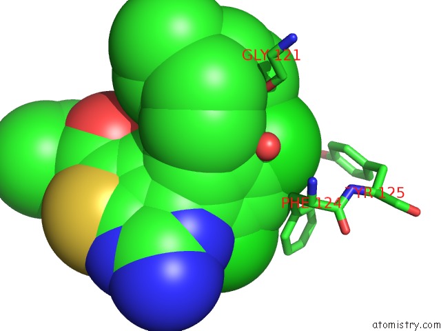
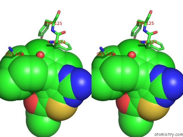
Chlorine binding site 1 out of 4 in 3omu
Go back to
Chlorine binding site 1 out
of 4 in the Crystal Structure of the N-Terminal Domain of An HSP90 From Trypanosoma Brucei, TB10.26.1080 in the Presence of A Thienopyrimidine Derivative
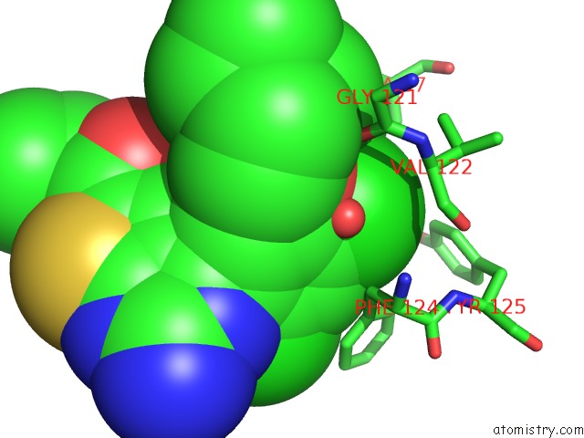
Mono view
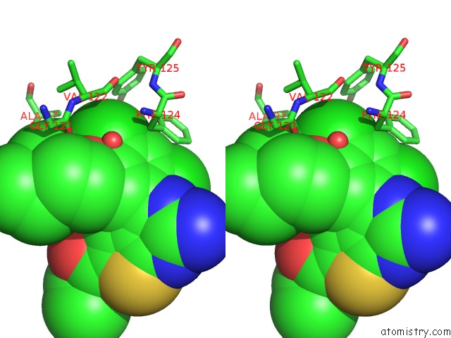
Stereo pair view
Mono view
Stereo pair view
A full contact list of Chlorine with other atoms in the Cl binding
site number 1 of Crystal Structure of the N-Terminal Domain of An HSP90 From Trypanosoma Brucei, TB10.26.1080 in the Presence of A Thienopyrimidine Derivative within 5.0Å range:
|
Chlorine binding site 2 out of 4 in 3omu
Go back to
Chlorine binding site 2 out
of 4 in the Crystal Structure of the N-Terminal Domain of An HSP90 From Trypanosoma Brucei, TB10.26.1080 in the Presence of A Thienopyrimidine Derivative
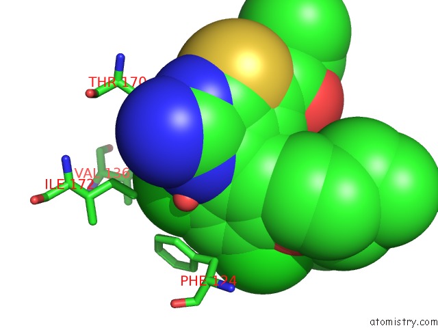
Mono view
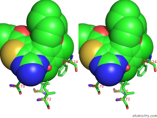
Stereo pair view
Mono view
Stereo pair view
A full contact list of Chlorine with other atoms in the Cl binding
site number 2 of Crystal Structure of the N-Terminal Domain of An HSP90 From Trypanosoma Brucei, TB10.26.1080 in the Presence of A Thienopyrimidine Derivative within 5.0Å range:
|
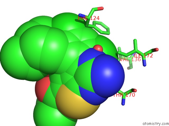
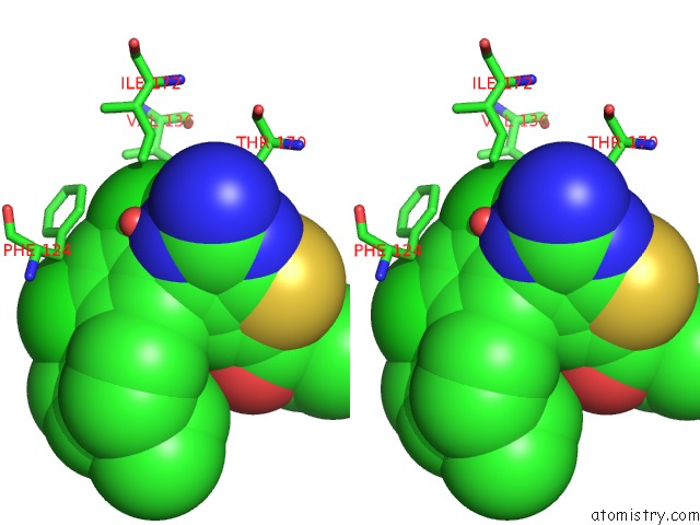
Chlorine binding site 3 out of 4 in 3omu
Go back to
Chlorine binding site 3 out
of 4 in the Crystal Structure of the N-Terminal Domain of An HSP90 From Trypanosoma Brucei, TB10.26.1080 in the Presence of A Thienopyrimidine Derivative
Mono view
Stereo pair view
Mono view
Stereo pair view
A full contact list of Chlorine with other atoms in the Cl binding
site number 3 of Crystal Structure of the N-Terminal Domain of An HSP90 From Trypanosoma Brucei, TB10.26.1080 in the Presence of A Thienopyrimidine Derivative within 5.0Å range:
|
Chlorine binding site 4 out of 4 in 3omu
Go back to
Chlorine binding site 4 out
of 4 in the Crystal Structure of the N-Terminal Domain of An HSP90 From Trypanosoma Brucei, TB10.26.1080 in the Presence of A Thienopyrimidine Derivative
Mono view
Stereo pair view
Mono view
Stereo pair view
A full contact list of Chlorine with other atoms in the Cl binding
site number 4 of Crystal Structure of the N-Terminal Domain of An HSP90 From Trypanosoma Brucei, TB10.26.1080 in the Presence of A Thienopyrimidine Derivative within 5.0Å range:
|
Reference:
J.C.Pizarro,
T.Hills,
G.Senisterra,
A.K.Wernimont,
C.Mackenzie,
N.R.Norcross,
M.A.Ferguson,
P.G.Wyatt,
I.H.Gilbert,
R.Hui.
Exploring the Trypanosoma Brucei HSP83 Potential As A Target For Structure Guided Drug Design. Plos Negl Trop Dis V. 7 E2492 2013.
ISSN: ESSN 1935-2735
PubMed: 24147171
DOI: 10.1371/JOURNAL.PNTD.0002492
Page generated: Sun Jul 21 01:48:02 2024
ISSN: ESSN 1935-2735
PubMed: 24147171
DOI: 10.1371/JOURNAL.PNTD.0002492
Last articles
Zn in 9MJ5Zn in 9HNW
Zn in 9G0L
Zn in 9FNE
Zn in 9DZN
Zn in 9E0I
Zn in 9D32
Zn in 9DAK
Zn in 8ZXC
Zn in 8ZUF