Chlorine »
PDB 3oox-3oxh »
3otg »
Chlorine in PDB 3otg: Crystal Structure of CALG1, Calicheamicin Glycostyltransferase, Tdp Bound Form
Protein crystallography data
The structure of Crystal Structure of CALG1, Calicheamicin Glycostyltransferase, Tdp Bound Form, PDB code: 3otg
was solved by
A.Chang,
S.Singh,
C.A.Bingman,
J.S.Thorson,
G.N.Phillips Jr.,
Center Foreukaryotic Structural Genomics (Cesg),
Enzyme Discovery For Naturalproduct Biosynthesis (Natpro),
with X-Ray Crystallography technique. A brief refinement statistics is given in the table below:
| Resolution Low / High (Å) | 45.98 / 2.08 |
| Space group | P 61 2 2 |
| Cell size a, b, c (Å), α, β, γ (°) | 106.177, 106.177, 155.817, 90.00, 90.00, 120.00 |
| R / Rfree (%) | 19.3 / 22.7 |
Chlorine Binding Sites:
The binding sites of Chlorine atom in the Crystal Structure of CALG1, Calicheamicin Glycostyltransferase, Tdp Bound Form
(pdb code 3otg). This binding sites where shown within
5.0 Angstroms radius around Chlorine atom.
In total 5 binding sites of Chlorine where determined in the Crystal Structure of CALG1, Calicheamicin Glycostyltransferase, Tdp Bound Form, PDB code: 3otg:
Jump to Chlorine binding site number: 1; 2; 3; 4; 5;
In total 5 binding sites of Chlorine where determined in the Crystal Structure of CALG1, Calicheamicin Glycostyltransferase, Tdp Bound Form, PDB code: 3otg:
Jump to Chlorine binding site number: 1; 2; 3; 4; 5;
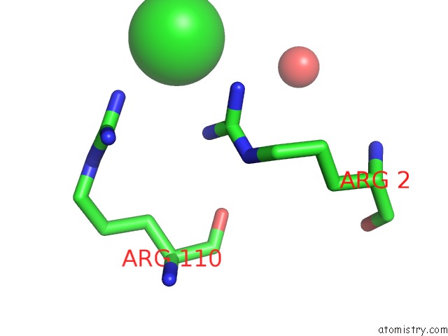
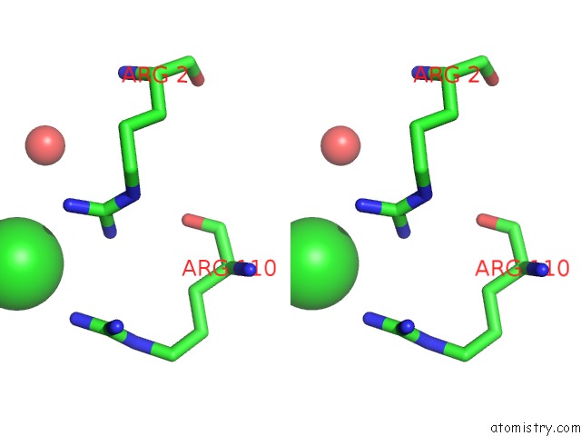
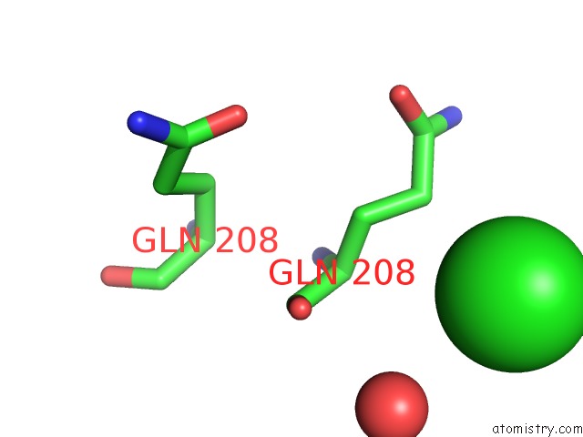
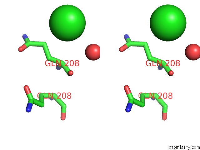
Chlorine binding site 1 out of 5 in 3otg
Go back to
Chlorine binding site 1 out
of 5 in the Crystal Structure of CALG1, Calicheamicin Glycostyltransferase, Tdp Bound Form
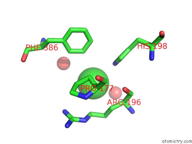
Mono view
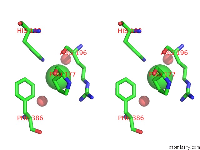
Stereo pair view
Mono view
Stereo pair view
A full contact list of Chlorine with other atoms in the Cl binding
site number 1 of Crystal Structure of CALG1, Calicheamicin Glycostyltransferase, Tdp Bound Form within 5.0Å range:
|
Chlorine binding site 2 out of 5 in 3otg
Go back to
Chlorine binding site 2 out
of 5 in the Crystal Structure of CALG1, Calicheamicin Glycostyltransferase, Tdp Bound Form
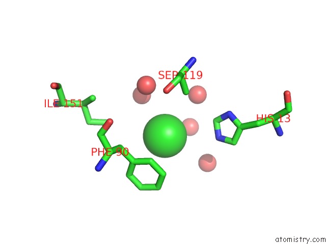
Mono view
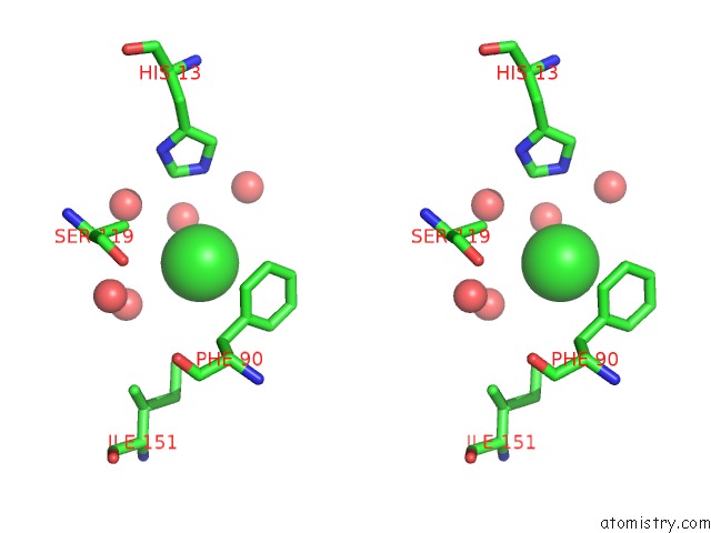
Stereo pair view
Mono view
Stereo pair view
A full contact list of Chlorine with other atoms in the Cl binding
site number 2 of Crystal Structure of CALG1, Calicheamicin Glycostyltransferase, Tdp Bound Form within 5.0Å range:
|
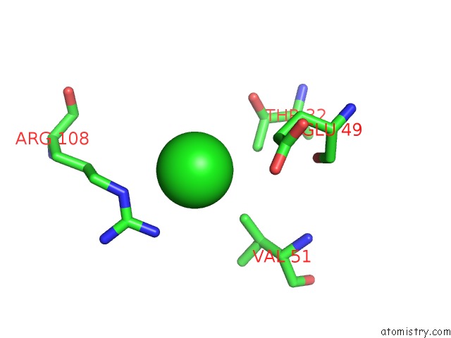
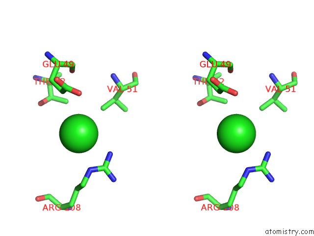
Chlorine binding site 3 out of 5 in 3otg
Go back to
Chlorine binding site 3 out
of 5 in the Crystal Structure of CALG1, Calicheamicin Glycostyltransferase, Tdp Bound Form
Mono view
Stereo pair view
Mono view
Stereo pair view
A full contact list of Chlorine with other atoms in the Cl binding
site number 3 of Crystal Structure of CALG1, Calicheamicin Glycostyltransferase, Tdp Bound Form within 5.0Å range:
|
Chlorine binding site 4 out of 5 in 3otg
Go back to
Chlorine binding site 4 out
of 5 in the Crystal Structure of CALG1, Calicheamicin Glycostyltransferase, Tdp Bound Form
Mono view
Stereo pair view
Mono view
Stereo pair view
A full contact list of Chlorine with other atoms in the Cl binding
site number 4 of Crystal Structure of CALG1, Calicheamicin Glycostyltransferase, Tdp Bound Form within 5.0Å range:
|
Chlorine binding site 5 out of 5 in 3otg
Go back to
Chlorine binding site 5 out
of 5 in the Crystal Structure of CALG1, Calicheamicin Glycostyltransferase, Tdp Bound Form
Mono view
Stereo pair view
Mono view
Stereo pair view
A full contact list of Chlorine with other atoms in the Cl binding
site number 5 of Crystal Structure of CALG1, Calicheamicin Glycostyltransferase, Tdp Bound Form within 5.0Å range:
|
Reference:
A.Chang,
S.Singh,
K.E.Helmich,
R.D.Goff,
C.A.Bingman,
J.S.Thorson,
G.N.Phillips.
Complete Set of Glycosyltransferase Structures in the Calicheamicin Biosynthetic Pathway Reveals the Origin of Regiospecificity. Proc.Natl.Acad.Sci.Usa V. 108 17649 2011.
ISSN: ISSN 0027-8424
PubMed: 21987796
DOI: 10.1073/PNAS.1108484108
Page generated: Sun Jul 21 01:56:36 2024
ISSN: ISSN 0027-8424
PubMed: 21987796
DOI: 10.1073/PNAS.1108484108
Last articles
Zn in 9MJ5Zn in 9HNW
Zn in 9G0L
Zn in 9FNE
Zn in 9DZN
Zn in 9E0I
Zn in 9D32
Zn in 9DAK
Zn in 8ZXC
Zn in 8ZUF