Chlorine »
PDB 3p4v-3pet »
3pd9 »
Chlorine in PDB 3pd9: X-Ray Structure of the Ligand-Binding Core of GLUA2 in Complex with (R)-5-Hpca at 2.1 A Resolution
Protein crystallography data
The structure of X-Ray Structure of the Ligand-Binding Core of GLUA2 in Complex with (R)-5-Hpca at 2.1 A Resolution, PDB code: 3pd9
was solved by
K.Frydenvang,
J.S.Kastrup,
with X-Ray Crystallography technique. A brief refinement statistics is given in the table below:
| Resolution Low / High (Å) | 29.42 / 2.10 |
| Space group | P 21 21 2 |
| Cell size a, b, c (Å), α, β, γ (°) | 99.391, 121.251, 47.547, 90.00, 90.00, 90.00 |
| R / Rfree (%) | 17.7 / 23.3 |
Chlorine Binding Sites:
The binding sites of Chlorine atom in the X-Ray Structure of the Ligand-Binding Core of GLUA2 in Complex with (R)-5-Hpca at 2.1 A Resolution
(pdb code 3pd9). This binding sites where shown within
5.0 Angstroms radius around Chlorine atom.
In total 4 binding sites of Chlorine where determined in the X-Ray Structure of the Ligand-Binding Core of GLUA2 in Complex with (R)-5-Hpca at 2.1 A Resolution, PDB code: 3pd9:
Jump to Chlorine binding site number: 1; 2; 3; 4;
In total 4 binding sites of Chlorine where determined in the X-Ray Structure of the Ligand-Binding Core of GLUA2 in Complex with (R)-5-Hpca at 2.1 A Resolution, PDB code: 3pd9:
Jump to Chlorine binding site number: 1; 2; 3; 4;
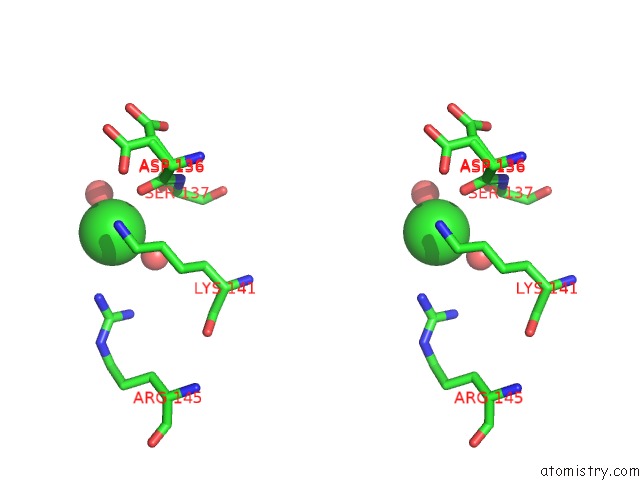
Chlorine binding site 1 out of 4 in 3pd9
Go back to
Chlorine binding site 1 out
of 4 in the X-Ray Structure of the Ligand-Binding Core of GLUA2 in Complex with (R)-5-Hpca at 2.1 A Resolution
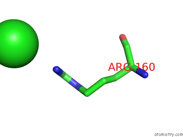
Mono view
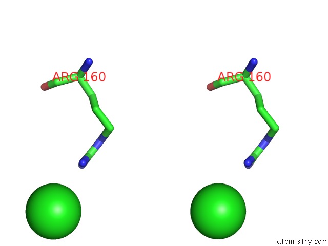
Stereo pair view
Mono view
Stereo pair view
A full contact list of Chlorine with other atoms in the Cl binding
site number 1 of X-Ray Structure of the Ligand-Binding Core of GLUA2 in Complex with (R)-5-Hpca at 2.1 A Resolution within 5.0Å range:
|
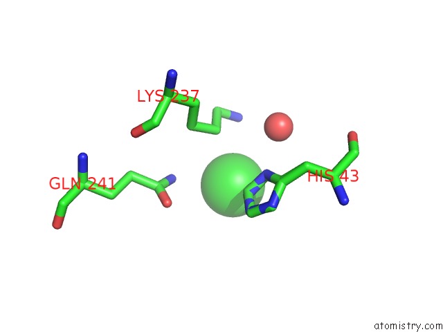
Chlorine binding site 2 out of 4 in 3pd9
Go back to
Chlorine binding site 2 out
of 4 in the X-Ray Structure of the Ligand-Binding Core of GLUA2 in Complex with (R)-5-Hpca at 2.1 A Resolution
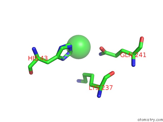
Mono view
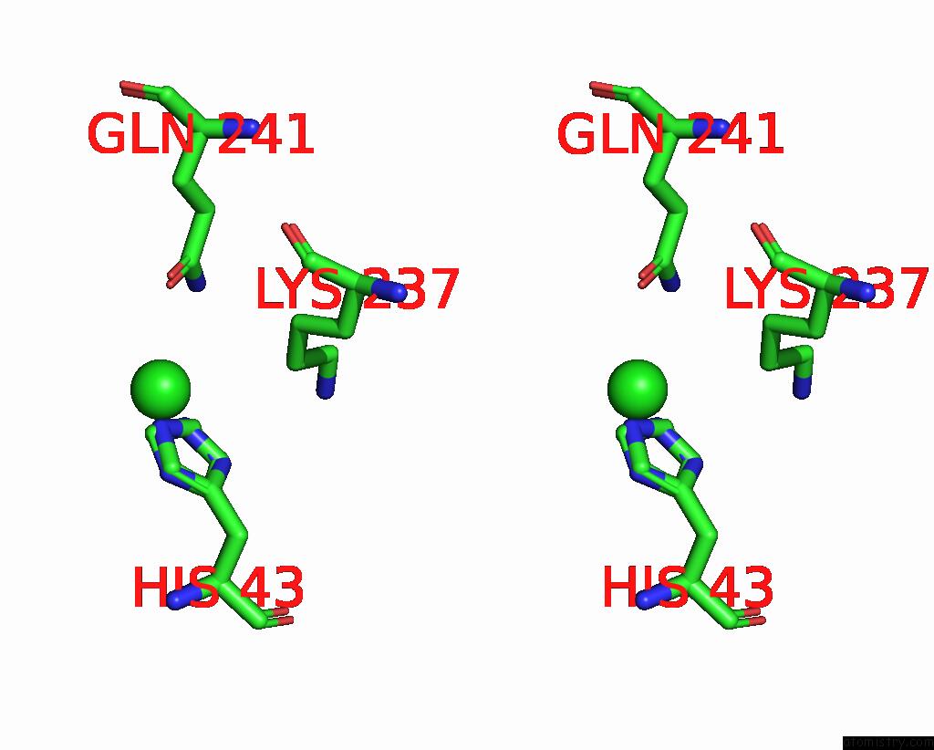
Stereo pair view
Mono view
Stereo pair view
A full contact list of Chlorine with other atoms in the Cl binding
site number 2 of X-Ray Structure of the Ligand-Binding Core of GLUA2 in Complex with (R)-5-Hpca at 2.1 A Resolution within 5.0Å range:
|
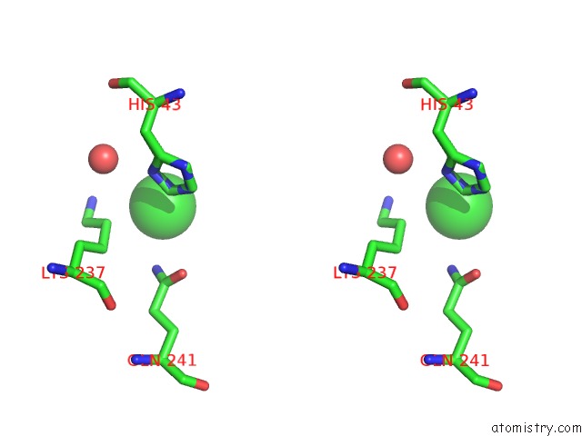
Chlorine binding site 3 out of 4 in 3pd9
Go back to
Chlorine binding site 3 out
of 4 in the X-Ray Structure of the Ligand-Binding Core of GLUA2 in Complex with (R)-5-Hpca at 2.1 A Resolution

Mono view
Stereo pair view
Mono view
Stereo pair view
A full contact list of Chlorine with other atoms in the Cl binding
site number 3 of X-Ray Structure of the Ligand-Binding Core of GLUA2 in Complex with (R)-5-Hpca at 2.1 A Resolution within 5.0Å range:
|
Chlorine binding site 4 out of 4 in 3pd9
Go back to
Chlorine binding site 4 out
of 4 in the X-Ray Structure of the Ligand-Binding Core of GLUA2 in Complex with (R)-5-Hpca at 2.1 A Resolution
Mono view
Stereo pair view
Mono view
Stereo pair view
A full contact list of Chlorine with other atoms in the Cl binding
site number 4 of X-Ray Structure of the Ligand-Binding Core of GLUA2 in Complex with (R)-5-Hpca at 2.1 A Resolution within 5.0Å range:
|
Reference:
K.Frydenvang,
D.S.Pickering,
J.R.Greenwood,
N.Krogsgaard-Larsen,
L.Brehm,
B.Nielsen,
S.B.Vogensen,
H.Hald,
J.S.Kastrup,
P.Krogsgaard-Larsen,
R.P.Clausen.
Biostructural and Pharmacological Studies of Bicyclic Analogues of the 3-Isoxazolol Glutamate Receptor Agonist Ibotenic Acid. J. Med. Chem. V. 53 8354 2010.
ISSN: ISSN 1520-4804
PubMed: 21067182
DOI: 10.1021/JM101218A
Page generated: Sun Jul 21 02:17:27 2024
ISSN: ISSN 1520-4804
PubMed: 21067182
DOI: 10.1021/JM101218A
Last articles
Zn in 9MJ5Zn in 9HNW
Zn in 9G0L
Zn in 9FNE
Zn in 9DZN
Zn in 9E0I
Zn in 9D32
Zn in 9DAK
Zn in 8ZXC
Zn in 8ZUF