Chlorine »
PDB 3rmn-3ruu »
3ruo »
Chlorine in PDB 3ruo: Complex Structure of Hevb EV93 Main Protease 3C with Rupintrivir (AG7088)
Enzymatic activity of Complex Structure of Hevb EV93 Main Protease 3C with Rupintrivir (AG7088)
All present enzymatic activity of Complex Structure of Hevb EV93 Main Protease 3C with Rupintrivir (AG7088):
3.4.22.28;
3.4.22.28;
Protein crystallography data
The structure of Complex Structure of Hevb EV93 Main Protease 3C with Rupintrivir (AG7088), PDB code: 3ruo
was solved by
Z.Kaczmarska,
R.Janowski,
L.Costenaro,
B.Coutard,
H.Norder,
B.Canard,
M.Coll,
with X-Ray Crystallography technique. A brief refinement statistics is given in the table below:
| Resolution Low / High (Å) | 30.00 / 1.50 |
| Space group | P 1 21 1 |
| Cell size a, b, c (Å), α, β, γ (°) | 38.995, 63.911, 66.357, 90.00, 90.43, 90.00 |
| R / Rfree (%) | 16.6 / 19.8 |
Other elements in 3ruo:
The structure of Complex Structure of Hevb EV93 Main Protease 3C with Rupintrivir (AG7088) also contains other interesting chemical elements:
| Fluorine | (F) | 2 atoms |
| Magnesium | (Mg) | 2 atoms |
Chlorine Binding Sites:
The binding sites of Chlorine atom in the Complex Structure of Hevb EV93 Main Protease 3C with Rupintrivir (AG7088)
(pdb code 3ruo). This binding sites where shown within
5.0 Angstroms radius around Chlorine atom.
In total only one binding site of Chlorine was determined in the Complex Structure of Hevb EV93 Main Protease 3C with Rupintrivir (AG7088), PDB code: 3ruo:
In total only one binding site of Chlorine was determined in the Complex Structure of Hevb EV93 Main Protease 3C with Rupintrivir (AG7088), PDB code: 3ruo:
Chlorine binding site 1 out of 1 in 3ruo
Go back to
Chlorine binding site 1 out
of 1 in the Complex Structure of Hevb EV93 Main Protease 3C with Rupintrivir (AG7088)
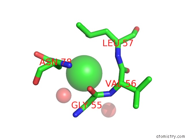
Mono view
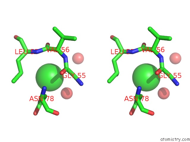
Stereo pair view
Mono view
Stereo pair view
A full contact list of Chlorine with other atoms in the Cl binding
site number 1 of Complex Structure of Hevb EV93 Main Protease 3C with Rupintrivir (AG7088) within 5.0Å range:
|
Reference:
L.Costenaro,
Z.Kaczmarska,
C.Arnan,
R.Janowski,
B.Coutard,
M.Sola,
A.E.Gorbalenya,
H.Norder,
B.Canard,
M.Coll.
Structural Basis For Antiviral Inhibition of the Main Protease, 3C, From Human Enterovirus 93. J.Virol. V. 85 10764 2011.
ISSN: ISSN 0022-538X
PubMed: 21835784
DOI: 10.1128/JVI.05062-11
Page generated: Sun Jul 21 03:59:15 2024
ISSN: ISSN 0022-538X
PubMed: 21835784
DOI: 10.1128/JVI.05062-11
Last articles
Zn in 9MJ5Zn in 9HNW
Zn in 9G0L
Zn in 9FNE
Zn in 9DZN
Zn in 9E0I
Zn in 9D32
Zn in 9DAK
Zn in 8ZXC
Zn in 8ZUF