Chlorine »
PDB 3rv4-3s8n »
3s0j »
Chlorine in PDB 3s0j: The Crystal Structure of Glycogen Phosphorylase B in Complex with 2,5- Dihydroxy-4-(Beta-D-Glucopyranosyl)-Chlorobenzene
Enzymatic activity of The Crystal Structure of Glycogen Phosphorylase B in Complex with 2,5- Dihydroxy-4-(Beta-D-Glucopyranosyl)-Chlorobenzene
All present enzymatic activity of The Crystal Structure of Glycogen Phosphorylase B in Complex with 2,5- Dihydroxy-4-(Beta-D-Glucopyranosyl)-Chlorobenzene:
2.4.1.1;
2.4.1.1;
Protein crystallography data
The structure of The Crystal Structure of Glycogen Phosphorylase B in Complex with 2,5- Dihydroxy-4-(Beta-D-Glucopyranosyl)-Chlorobenzene, PDB code: 3s0j
was solved by
K.-M.Alexacou,
S.E.Zographos,
E.D.Chrysina,
N.G.Oikonomakos,
D.D.Leonidas,
with X-Ray Crystallography technique. A brief refinement statistics is given in the table below:
| Resolution Low / High (Å) | 30.00 / 2.00 |
| Space group | P 43 21 2 |
| Cell size a, b, c (Å), α, β, γ (°) | 128.450, 128.450, 116.364, 90.00, 90.00, 90.00 |
| R / Rfree (%) | 19 / 20.9 |
Chlorine Binding Sites:
The binding sites of Chlorine atom in the The Crystal Structure of Glycogen Phosphorylase B in Complex with 2,5- Dihydroxy-4-(Beta-D-Glucopyranosyl)-Chlorobenzene
(pdb code 3s0j). This binding sites where shown within
5.0 Angstroms radius around Chlorine atom.
In total only one binding site of Chlorine was determined in the The Crystal Structure of Glycogen Phosphorylase B in Complex with 2,5- Dihydroxy-4-(Beta-D-Glucopyranosyl)-Chlorobenzene, PDB code: 3s0j:
In total only one binding site of Chlorine was determined in the The Crystal Structure of Glycogen Phosphorylase B in Complex with 2,5- Dihydroxy-4-(Beta-D-Glucopyranosyl)-Chlorobenzene, PDB code: 3s0j:
Chlorine binding site 1 out of 1 in 3s0j
Go back to
Chlorine binding site 1 out
of 1 in the The Crystal Structure of Glycogen Phosphorylase B in Complex with 2,5- Dihydroxy-4-(Beta-D-Glucopyranosyl)-Chlorobenzene
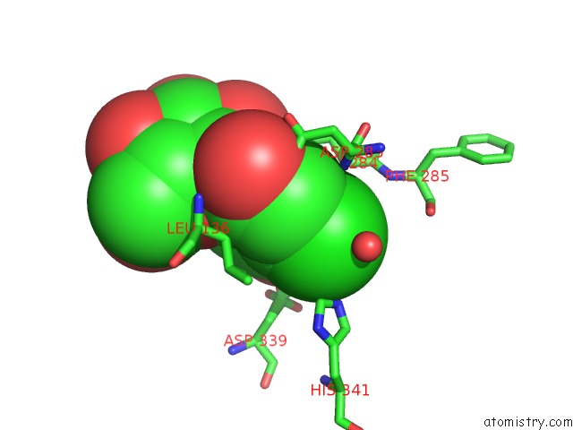
Mono view
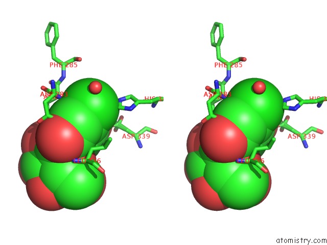
Stereo pair view
Mono view
Stereo pair view
A full contact list of Chlorine with other atoms in the Cl binding
site number 1 of The Crystal Structure of Glycogen Phosphorylase B in Complex with 2,5- Dihydroxy-4-(Beta-D-Glucopyranosyl)-Chlorobenzene within 5.0Å range:
|
Reference:
K.M.Alexacou,
Y.Z.Zhang,
J.P.Praly,
S.E.Zographos,
E.D.Chrysina,
N.G.Oikonomakos,
D.D.Leonidas.
Halogen-Substituted (C-Beta-D-Glucopyranosyl)-Hydroquinone Regioisomers: Synthesis, Enzymatic Evaluation and Their Binding to Glycogen Phosphorylase. Bioorg.Med.Chem. V. 19 5125 2011.
ISSN: ISSN 0968-0896
PubMed: 21821421
DOI: 10.1016/J.BMC.2011.07.024
Page generated: Fri Jul 11 10:04:35 2025
ISSN: ISSN 0968-0896
PubMed: 21821421
DOI: 10.1016/J.BMC.2011.07.024
Last articles
Cl in 4HCJCl in 4HB4
Cl in 4HBU
Cl in 4HBT
Cl in 4HB2
Cl in 4HAZ
Cl in 4HB0
Cl in 4HB3
Cl in 4HAY
Cl in 4HAX