Chlorine »
PDB 3rv4-3s8n »
3s18 »
Chlorine in PDB 3s18: Crystal Structure of A Plant Albumin From Cicer Arietinum Showing Hemagglutination
Protein crystallography data
The structure of Crystal Structure of A Plant Albumin From Cicer Arietinum Showing Hemagglutination, PDB code: 3s18
was solved by
U.Sharma,
C.G.Suresh,
with X-Ray Crystallography technique. A brief refinement statistics is given in the table below:
| Resolution Low / High (Å) | 35.30 / 2.20 |
| Space group | P 3 |
| Cell size a, b, c (Å), α, β, γ (°) | 81.917, 81.917, 69.591, 90.00, 90.00, 120.00 |
| R / Rfree (%) | 17.7 / 22.3 |
Other elements in 3s18:
The structure of Crystal Structure of A Plant Albumin From Cicer Arietinum Showing Hemagglutination also contains other interesting chemical elements:
| Iodine | (I) | 2 atoms |
| Calcium | (Ca) | 2 atoms |
| Sodium | (Na) | 1 atom |
Chlorine Binding Sites:
The binding sites of Chlorine atom in the Crystal Structure of A Plant Albumin From Cicer Arietinum Showing Hemagglutination
(pdb code 3s18). This binding sites where shown within
5.0 Angstroms radius around Chlorine atom.
In total 3 binding sites of Chlorine where determined in the Crystal Structure of A Plant Albumin From Cicer Arietinum Showing Hemagglutination, PDB code: 3s18:
Jump to Chlorine binding site number: 1; 2; 3;
In total 3 binding sites of Chlorine where determined in the Crystal Structure of A Plant Albumin From Cicer Arietinum Showing Hemagglutination, PDB code: 3s18:
Jump to Chlorine binding site number: 1; 2; 3;
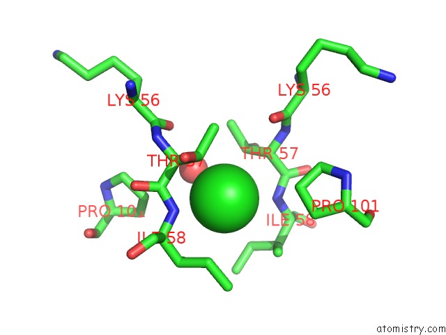
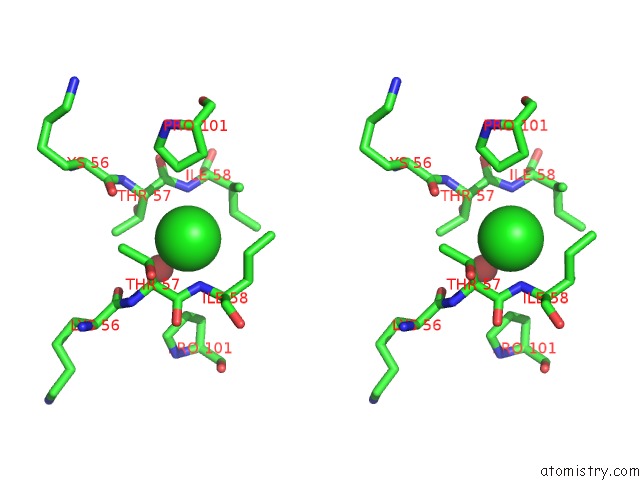
Chlorine binding site 1 out of 3 in 3s18
Go back to
Chlorine binding site 1 out
of 3 in the Crystal Structure of A Plant Albumin From Cicer Arietinum Showing Hemagglutination
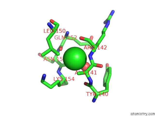
Mono view
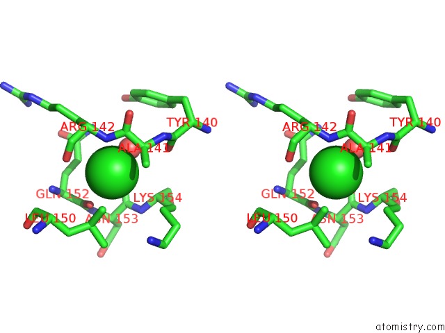
Stereo pair view
Mono view
Stereo pair view
A full contact list of Chlorine with other atoms in the Cl binding
site number 1 of Crystal Structure of A Plant Albumin From Cicer Arietinum Showing Hemagglutination within 5.0Å range:
|
Chlorine binding site 2 out of 3 in 3s18
Go back to
Chlorine binding site 2 out
of 3 in the Crystal Structure of A Plant Albumin From Cicer Arietinum Showing Hemagglutination
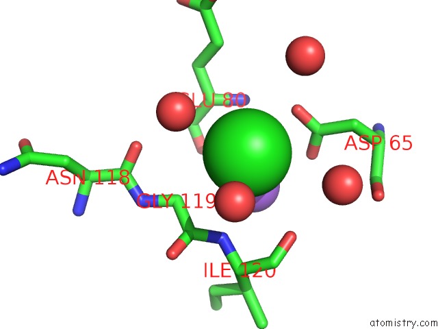
Mono view
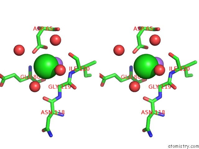
Stereo pair view
Mono view
Stereo pair view
A full contact list of Chlorine with other atoms in the Cl binding
site number 2 of Crystal Structure of A Plant Albumin From Cicer Arietinum Showing Hemagglutination within 5.0Å range:
|
Chlorine binding site 3 out of 3 in 3s18
Go back to
Chlorine binding site 3 out
of 3 in the Crystal Structure of A Plant Albumin From Cicer Arietinum Showing Hemagglutination
Mono view
Stereo pair view
Mono view
Stereo pair view
A full contact list of Chlorine with other atoms in the Cl binding
site number 3 of Crystal Structure of A Plant Albumin From Cicer Arietinum Showing Hemagglutination within 5.0Å range:
|
Reference:
U.Sharma,
U.V.Katre,
C.G.Suresh.
Crystal Structure of A Plant Albumin From Cicer Arietinum (Chickpea) Possessing Hemopexin Fold and Hemagglutination Activity Planta 2015.
ISSN: ISSN 0032-0935
PubMed: 25559942
DOI: 10.1007/S00425-014-2236-6
Page generated: Sun Jul 21 04:04:38 2024
ISSN: ISSN 0032-0935
PubMed: 25559942
DOI: 10.1007/S00425-014-2236-6
Last articles
Zn in 9J0NZn in 9J0O
Zn in 9J0P
Zn in 9FJX
Zn in 9EKB
Zn in 9C0F
Zn in 9CAH
Zn in 9CH0
Zn in 9CH3
Zn in 9CH1