Chlorine »
PDB 3s8o-3seo »
3s8w »
Chlorine in PDB 3s8w: D2 Domain of Human IFNAR2
Protein crystallography data
The structure of D2 Domain of Human IFNAR2, PDB code: 3s8w
was solved by
C.Thomas,
K.C.Garcia,
with X-Ray Crystallography technique. A brief refinement statistics is given in the table below:
| Resolution Low / High (Å) | 19.93 / 2.60 |
| Space group | P 65 2 2 |
| Cell size a, b, c (Å), α, β, γ (°) | 82.660, 82.660, 225.330, 90.00, 90.00, 120.00 |
| R / Rfree (%) | 21.8 / 27 |
Chlorine Binding Sites:
The binding sites of Chlorine atom in the D2 Domain of Human IFNAR2
(pdb code 3s8w). This binding sites where shown within
5.0 Angstroms radius around Chlorine atom.
In total 2 binding sites of Chlorine where determined in the D2 Domain of Human IFNAR2, PDB code: 3s8w:
Jump to Chlorine binding site number: 1; 2;
In total 2 binding sites of Chlorine where determined in the D2 Domain of Human IFNAR2, PDB code: 3s8w:
Jump to Chlorine binding site number: 1; 2;
Chlorine binding site 1 out of 2 in 3s8w
Go back to
Chlorine binding site 1 out
of 2 in the D2 Domain of Human IFNAR2
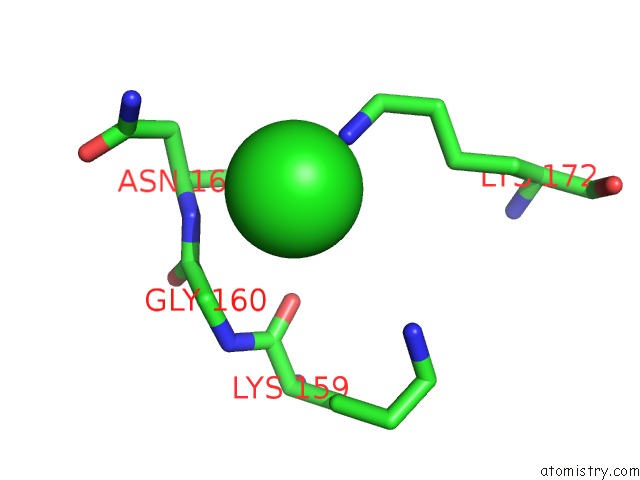
Mono view
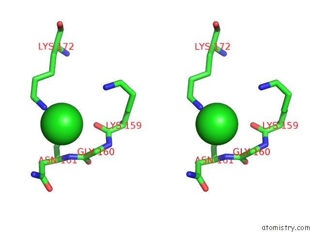
Stereo pair view
Mono view
Stereo pair view
A full contact list of Chlorine with other atoms in the Cl binding
site number 1 of D2 Domain of Human IFNAR2 within 5.0Å range:
|
Chlorine binding site 2 out of 2 in 3s8w
Go back to
Chlorine binding site 2 out
of 2 in the D2 Domain of Human IFNAR2
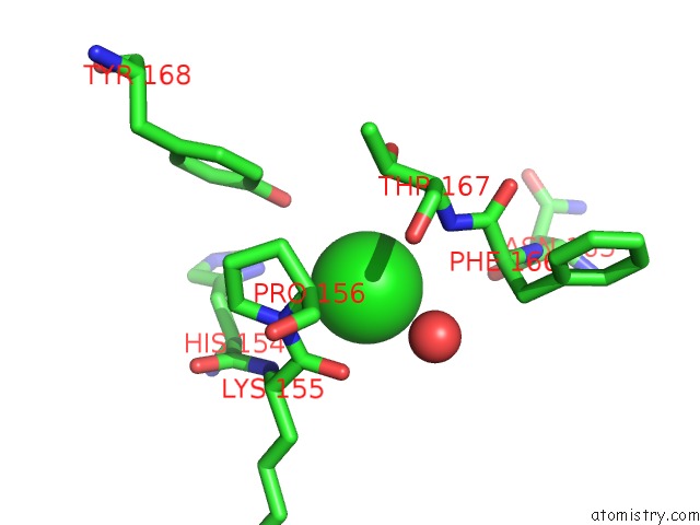
Mono view
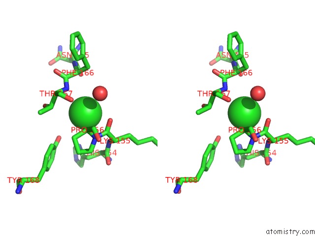
Stereo pair view
Mono view
Stereo pair view
A full contact list of Chlorine with other atoms in the Cl binding
site number 2 of D2 Domain of Human IFNAR2 within 5.0Å range:
|
Reference:
C.Thomas,
I.Moraga,
D.Levin,
P.O.Krutzik,
Y.Podoplelova,
A.Trejo,
C.Lee,
G.Yarden,
S.E.Vleck,
J.S.Glenn,
G.P.Nolan,
J.Piehler,
G.Schreiber,
K.C.Garcia.
Structural Linkage Between Ligand Discrimination and Receptor Activation By Type I Interferons. Cell(Cambridge,Mass.) V. 146 621 2011.
ISSN: ISSN 0092-8674
PubMed: 21854986
DOI: 10.1016/J.CELL.2011.06.048
Page generated: Sun Jul 21 04:14:15 2024
ISSN: ISSN 0092-8674
PubMed: 21854986
DOI: 10.1016/J.CELL.2011.06.048
Last articles
Zn in 9J0NZn in 9J0O
Zn in 9J0P
Zn in 9FJX
Zn in 9EKB
Zn in 9C0F
Zn in 9CAH
Zn in 9CH0
Zn in 9CH3
Zn in 9CH1