Chlorine »
PDB 3s8o-3seo »
3sd7 »
Chlorine in PDB 3sd7: 1.7 Angstrom Resolution Crystal Structure of Putative Phosphatase From Clostridium Difficile
Protein crystallography data
The structure of 1.7 Angstrom Resolution Crystal Structure of Putative Phosphatase From Clostridium Difficile, PDB code: 3sd7
was solved by
G.Minasov,
L.Shuvalova,
I.Dubrovska,
J.Winsor,
L.Papazisi,
W.F.Anderson,
Center For Structural Genomics Of Infectious Diseases (Csgid),
with X-Ray Crystallography technique. A brief refinement statistics is given in the table below:
| Resolution Low / High (Å) | 29.65 / 1.70 |
| Space group | I 41 |
| Cell size a, b, c (Å), α, β, γ (°) | 116.333, 116.333, 36.080, 90.00, 90.00, 90.00 |
| R / Rfree (%) | 16.7 / 19.3 |
Other elements in 3sd7:
The structure of 1.7 Angstrom Resolution Crystal Structure of Putative Phosphatase From Clostridium Difficile also contains other interesting chemical elements:
| Sodium | (Na) | 1 atom |
Chlorine Binding Sites:
The binding sites of Chlorine atom in the 1.7 Angstrom Resolution Crystal Structure of Putative Phosphatase From Clostridium Difficile
(pdb code 3sd7). This binding sites where shown within
5.0 Angstroms radius around Chlorine atom.
In total only one binding site of Chlorine was determined in the 1.7 Angstrom Resolution Crystal Structure of Putative Phosphatase From Clostridium Difficile, PDB code: 3sd7:
In total only one binding site of Chlorine was determined in the 1.7 Angstrom Resolution Crystal Structure of Putative Phosphatase From Clostridium Difficile, PDB code: 3sd7:
Chlorine binding site 1 out of 1 in 3sd7
Go back to
Chlorine binding site 1 out
of 1 in the 1.7 Angstrom Resolution Crystal Structure of Putative Phosphatase From Clostridium Difficile
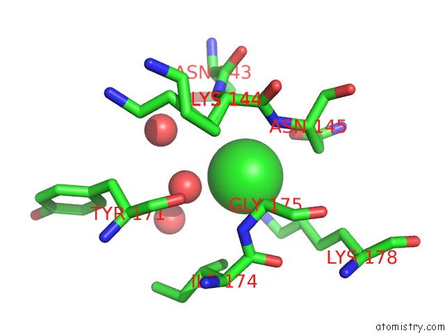
Mono view
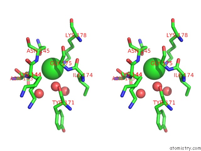
Stereo pair view
Mono view
Stereo pair view
A full contact list of Chlorine with other atoms in the Cl binding
site number 1 of 1.7 Angstrom Resolution Crystal Structure of Putative Phosphatase From Clostridium Difficile within 5.0Å range:
|
Reference:
G.Minasov,
L.Shuvalova,
I.Dubrovska,
J.Winsor,
L.Papazisi,
W.F.Anderson,
Center For Structural Genomics Of Infectious Diseases(Csgid).
1.7 Angstrom Resolution Crystal Structure of Putative Phosphatase From Clostridium Difficile. To Be Published.
Page generated: Sun Jul 21 04:21:41 2024
Last articles
Ca in 5O5TCa in 5O5R
Ca in 5NZF
Ca in 5O2Z
Ca in 5O2Y
Ca in 5O0Z
Ca in 5O25
Ca in 5O1U
Ca in 5O0S
Ca in 5NZE