Chlorine »
PDB 3ser-3smc »
3sg9 »
Chlorine in PDB 3sg9: Crystal Structure of Aminoglycoside-2''-Phosphotransferase Type Iva Kanamycin A Complex
Protein crystallography data
The structure of Crystal Structure of Aminoglycoside-2''-Phosphotransferase Type Iva Kanamycin A Complex, PDB code: 3sg9
was solved by
K.Shi,
D.R.Houston,
A.M.Berghuis,
with X-Ray Crystallography technique. A brief refinement statistics is given in the table below:
| Resolution Low / High (Å) | 50.00 / 2.15 |
| Space group | P 1 21 1 |
| Cell size a, b, c (Å), α, β, γ (°) | 42.903, 101.426, 73.399, 90.00, 100.61, 90.00 |
| R / Rfree (%) | 19.5 / 25.1 |
Chlorine Binding Sites:
The binding sites of Chlorine atom in the Crystal Structure of Aminoglycoside-2''-Phosphotransferase Type Iva Kanamycin A Complex
(pdb code 3sg9). This binding sites where shown within
5.0 Angstroms radius around Chlorine atom.
In total only one binding site of Chlorine was determined in the Crystal Structure of Aminoglycoside-2''-Phosphotransferase Type Iva Kanamycin A Complex, PDB code: 3sg9:
In total only one binding site of Chlorine was determined in the Crystal Structure of Aminoglycoside-2''-Phosphotransferase Type Iva Kanamycin A Complex, PDB code: 3sg9:
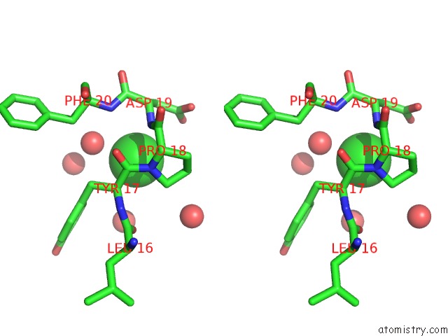
Chlorine binding site 1 out of 1 in 3sg9
Go back to
Chlorine binding site 1 out
of 1 in the Crystal Structure of Aminoglycoside-2''-Phosphotransferase Type Iva Kanamycin A Complex
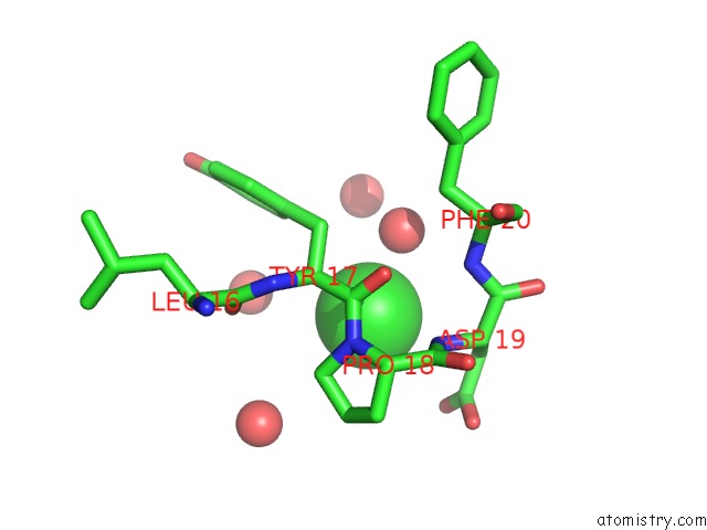
Mono view
Stereo pair view
Mono view
Stereo pair view
A full contact list of Chlorine with other atoms in the Cl binding
site number 1 of Crystal Structure of Aminoglycoside-2''-Phosphotransferase Type Iva Kanamycin A Complex within 5.0Å range:
|
Reference:
K.Shi,
D.R.Houston,
A.M.Berghuis.
Crystal Structures of Antibiotic-Bound Complexes of Aminoglycoside 2''-Phosphotransferase Iva Highlight the Diversity in Substrate Binding Modes Among Aminoglycoside Kinases. Biochemistry V. 50 6237 2011.
ISSN: ISSN 0006-2960
PubMed: 21678960
DOI: 10.1021/BI200747F
Page generated: Sun Jul 21 04:29:19 2024
ISSN: ISSN 0006-2960
PubMed: 21678960
DOI: 10.1021/BI200747F
Last articles
Zn in 9J0NZn in 9J0O
Zn in 9J0P
Zn in 9FJX
Zn in 9EKB
Zn in 9C0F
Zn in 9CAH
Zn in 9CH0
Zn in 9CH3
Zn in 9CH1