Chlorine »
PDB 3sml-3ste »
3sns »
Chlorine in PDB 3sns: Crystal Structure of the C-Terminal Domain of Escherichia Coli Lipoprotein Bamc
Protein crystallography data
The structure of Crystal Structure of the C-Terminal Domain of Escherichia Coli Lipoprotein Bamc, PDB code: 3sns
was solved by
K.H.Kim,
S.Aulakh,
W.Tan,
M.Paetzel,
with X-Ray Crystallography technique. A brief refinement statistics is given in the table below:
| Resolution Low / High (Å) | 41.82 / 1.50 |
| Space group | H 3 |
| Cell size a, b, c (Å), α, β, γ (°) | 78.850, 78.850, 52.900, 90.00, 90.00, 120.00 |
| R / Rfree (%) | 16.1 / 18.3 |
Chlorine Binding Sites:
The binding sites of Chlorine atom in the Crystal Structure of the C-Terminal Domain of Escherichia Coli Lipoprotein Bamc
(pdb code 3sns). This binding sites where shown within
5.0 Angstroms radius around Chlorine atom.
In total 2 binding sites of Chlorine where determined in the Crystal Structure of the C-Terminal Domain of Escherichia Coli Lipoprotein Bamc, PDB code: 3sns:
Jump to Chlorine binding site number: 1; 2;
In total 2 binding sites of Chlorine where determined in the Crystal Structure of the C-Terminal Domain of Escherichia Coli Lipoprotein Bamc, PDB code: 3sns:
Jump to Chlorine binding site number: 1; 2;
Chlorine binding site 1 out of 2 in 3sns
Go back to
Chlorine binding site 1 out
of 2 in the Crystal Structure of the C-Terminal Domain of Escherichia Coli Lipoprotein Bamc

Mono view
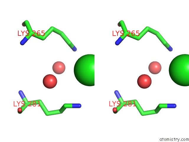
Stereo pair view
Mono view
Stereo pair view
A full contact list of Chlorine with other atoms in the Cl binding
site number 1 of Crystal Structure of the C-Terminal Domain of Escherichia Coli Lipoprotein Bamc within 5.0Å range:
|
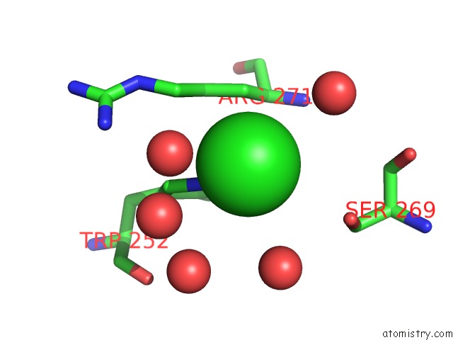
Chlorine binding site 2 out of 2 in 3sns
Go back to
Chlorine binding site 2 out
of 2 in the Crystal Structure of the C-Terminal Domain of Escherichia Coli Lipoprotein Bamc
Mono view

Stereo pair view
Mono view
Stereo pair view
A full contact list of Chlorine with other atoms in the Cl binding
site number 2 of Crystal Structure of the C-Terminal Domain of Escherichia Coli Lipoprotein Bamc within 5.0Å range:
|
Reference:
K.H.Kim,
S.Aulakh,
W.Tan,
M.Paetzel.
Crystallographic Analysis of the C-Terminal Domain of the Escherichia Coli Lipoprotein Bamc. Acta Crystallogr.,Sect.F V. 67 1350 2011.
ISSN: ESSN 1744-3091
PubMed: 22102230
DOI: 10.1107/S174430911103363X
Page generated: Sun Jul 21 04:40:49 2024
ISSN: ESSN 1744-3091
PubMed: 22102230
DOI: 10.1107/S174430911103363X
Last articles
Zn in 9J0NZn in 9J0O
Zn in 9J0P
Zn in 9FJX
Zn in 9EKB
Zn in 9C0F
Zn in 9CAH
Zn in 9CH0
Zn in 9CH3
Zn in 9CH1