Chlorine »
PDB 3sml-3ste »
3st5 »
Chlorine in PDB 3st5: Crystal Structure of Wild-Type Hiv-1 Protease with C3-Substituted Hexahydrocyclopentafuranyl Urethane As P2-Ligand, Grl-0489A
Enzymatic activity of Crystal Structure of Wild-Type Hiv-1 Protease with C3-Substituted Hexahydrocyclopentafuranyl Urethane As P2-Ligand, Grl-0489A
All present enzymatic activity of Crystal Structure of Wild-Type Hiv-1 Protease with C3-Substituted Hexahydrocyclopentafuranyl Urethane As P2-Ligand, Grl-0489A:
3.4.23.16;
3.4.23.16;
Protein crystallography data
The structure of Crystal Structure of Wild-Type Hiv-1 Protease with C3-Substituted Hexahydrocyclopentafuranyl Urethane As P2-Ligand, Grl-0489A, PDB code: 3st5
was solved by
Y.-F.Wang,
J.Agniswamy,
I.T.Weber,
with X-Ray Crystallography technique. A brief refinement statistics is given in the table below:
| Resolution Low / High (Å) | 10.00 / 1.45 |
| Space group | P 21 21 2 |
| Cell size a, b, c (Å), α, β, γ (°) | 58.390, 86.552, 45.846, 90.00, 90.00, 90.00 |
| R / Rfree (%) | 15.7 / 21.9 |
Chlorine Binding Sites:
The binding sites of Chlorine atom in the Crystal Structure of Wild-Type Hiv-1 Protease with C3-Substituted Hexahydrocyclopentafuranyl Urethane As P2-Ligand, Grl-0489A
(pdb code 3st5). This binding sites where shown within
5.0 Angstroms radius around Chlorine atom.
In total 2 binding sites of Chlorine where determined in the Crystal Structure of Wild-Type Hiv-1 Protease with C3-Substituted Hexahydrocyclopentafuranyl Urethane As P2-Ligand, Grl-0489A, PDB code: 3st5:
Jump to Chlorine binding site number: 1; 2;
In total 2 binding sites of Chlorine where determined in the Crystal Structure of Wild-Type Hiv-1 Protease with C3-Substituted Hexahydrocyclopentafuranyl Urethane As P2-Ligand, Grl-0489A, PDB code: 3st5:
Jump to Chlorine binding site number: 1; 2;
Chlorine binding site 1 out of 2 in 3st5
Go back to
Chlorine binding site 1 out
of 2 in the Crystal Structure of Wild-Type Hiv-1 Protease with C3-Substituted Hexahydrocyclopentafuranyl Urethane As P2-Ligand, Grl-0489A
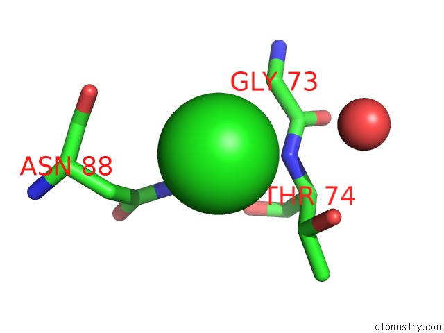
Mono view
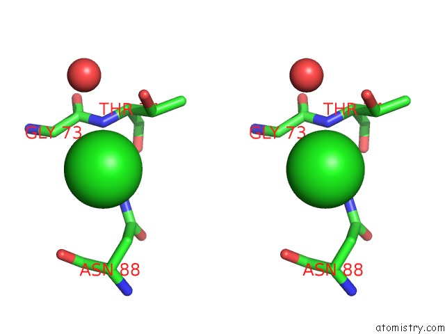
Stereo pair view
Mono view
Stereo pair view
A full contact list of Chlorine with other atoms in the Cl binding
site number 1 of Crystal Structure of Wild-Type Hiv-1 Protease with C3-Substituted Hexahydrocyclopentafuranyl Urethane As P2-Ligand, Grl-0489A within 5.0Å range:
|
Chlorine binding site 2 out of 2 in 3st5
Go back to
Chlorine binding site 2 out
of 2 in the Crystal Structure of Wild-Type Hiv-1 Protease with C3-Substituted Hexahydrocyclopentafuranyl Urethane As P2-Ligand, Grl-0489A
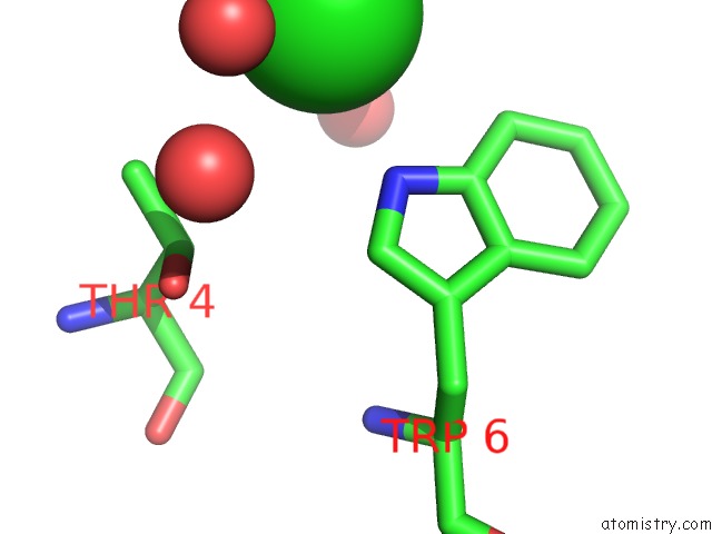
Mono view
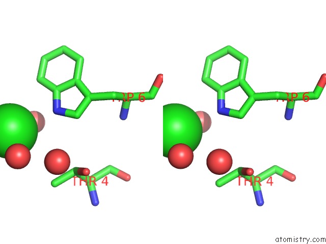
Stereo pair view
Mono view
Stereo pair view
A full contact list of Chlorine with other atoms in the Cl binding
site number 2 of Crystal Structure of Wild-Type Hiv-1 Protease with C3-Substituted Hexahydrocyclopentafuranyl Urethane As P2-Ligand, Grl-0489A within 5.0Å range:
|
Reference:
A.K.Ghosh,
B.D.Chapsal,
G.L.Parham,
M.Steffey,
J.Agniswamy,
Y.F.Wang,
M.Amano,
I.T.Weber,
H.Mitsuya.
Design of Hiv-1 Protease Inhibitors with C3-Substituted Hexahydrocyclopentafuranyl Urethanes As P2-Ligands: Synthesis, Biological Evaluation, and Protein-Ligand X-Ray Crystal Structure. J.Med.Chem. V. 54 5890 2011.
ISSN: ISSN 0022-2623
PubMed: 21800876
DOI: 10.1021/JM200649P
Page generated: Sun Jul 21 04:46:49 2024
ISSN: ISSN 0022-2623
PubMed: 21800876
DOI: 10.1021/JM200649P
Last articles
Zn in 9MJ5Zn in 9HNW
Zn in 9G0L
Zn in 9FNE
Zn in 9DZN
Zn in 9E0I
Zn in 9D32
Zn in 9DAK
Zn in 8ZXC
Zn in 8ZUF