Chlorine »
PDB 3stf-3t4u »
3t1a »
Chlorine in PDB 3t1a: Crystal Structure of Hiv-1 Reverse Transcriptase (K103N Mutant) in Complex with Inhibitor M05
Enzymatic activity of Crystal Structure of Hiv-1 Reverse Transcriptase (K103N Mutant) in Complex with Inhibitor M05
All present enzymatic activity of Crystal Structure of Hiv-1 Reverse Transcriptase (K103N Mutant) in Complex with Inhibitor M05:
2.7.7.49; 2.7.7.7; 3.1.26.13;
2.7.7.49; 2.7.7.7; 3.1.26.13;
Protein crystallography data
The structure of Crystal Structure of Hiv-1 Reverse Transcriptase (K103N Mutant) in Complex with Inhibitor M05, PDB code: 3t1a
was solved by
Y.Yan,
J.Reid,
with X-Ray Crystallography technique. A brief refinement statistics is given in the table below:
| Resolution Low / High (Å) | 35.14 / 2.40 |
| Space group | C 2 2 21 |
| Cell size a, b, c (Å), α, β, γ (°) | 118.330, 154.230, 155.680, 90.00, 90.00, 90.00 |
| R / Rfree (%) | 20.5 / 25 |
Chlorine Binding Sites:
The binding sites of Chlorine atom in the Crystal Structure of Hiv-1 Reverse Transcriptase (K103N Mutant) in Complex with Inhibitor M05
(pdb code 3t1a). This binding sites where shown within
5.0 Angstroms radius around Chlorine atom.
In total 3 binding sites of Chlorine where determined in the Crystal Structure of Hiv-1 Reverse Transcriptase (K103N Mutant) in Complex with Inhibitor M05, PDB code: 3t1a:
Jump to Chlorine binding site number: 1; 2; 3;
In total 3 binding sites of Chlorine where determined in the Crystal Structure of Hiv-1 Reverse Transcriptase (K103N Mutant) in Complex with Inhibitor M05, PDB code: 3t1a:
Jump to Chlorine binding site number: 1; 2; 3;
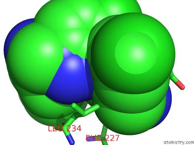
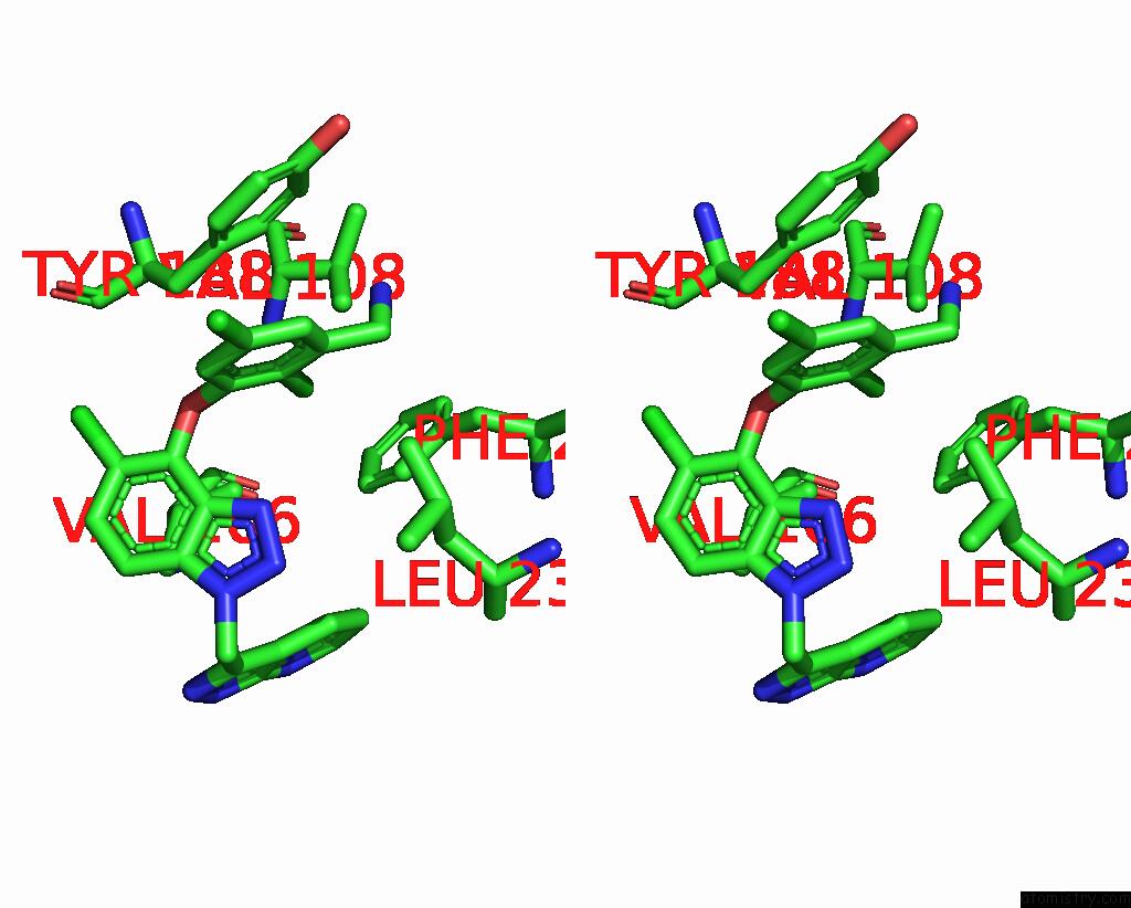
Chlorine binding site 1 out of 3 in 3t1a
Go back to
Chlorine binding site 1 out
of 3 in the Crystal Structure of Hiv-1 Reverse Transcriptase (K103N Mutant) in Complex with Inhibitor M05
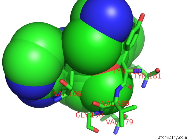
Mono view
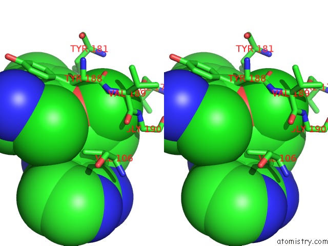
Stereo pair view
Mono view
Stereo pair view
A full contact list of Chlorine with other atoms in the Cl binding
site number 1 of Crystal Structure of Hiv-1 Reverse Transcriptase (K103N Mutant) in Complex with Inhibitor M05 within 5.0Å range:
|
Chlorine binding site 2 out of 3 in 3t1a
Go back to
Chlorine binding site 2 out
of 3 in the Crystal Structure of Hiv-1 Reverse Transcriptase (K103N Mutant) in Complex with Inhibitor M05
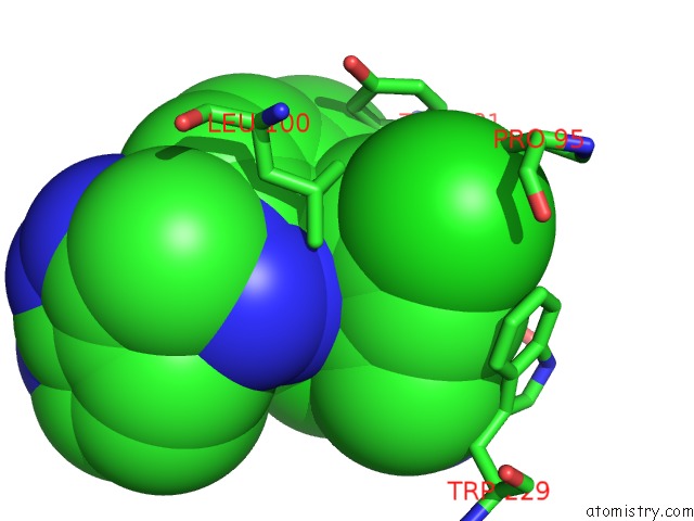
Mono view
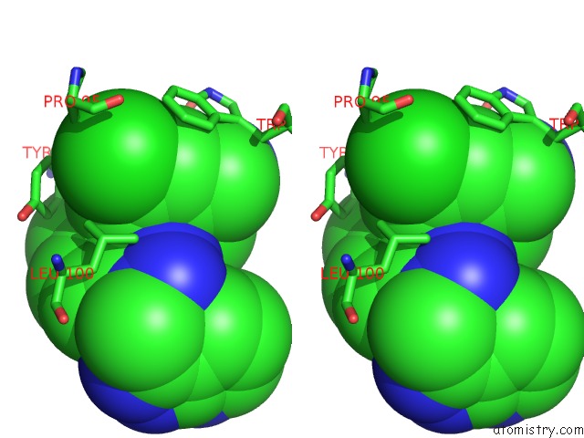
Stereo pair view
Mono view
Stereo pair view
A full contact list of Chlorine with other atoms in the Cl binding
site number 2 of Crystal Structure of Hiv-1 Reverse Transcriptase (K103N Mutant) in Complex with Inhibitor M05 within 5.0Å range:
|
Chlorine binding site 3 out of 3 in 3t1a
Go back to
Chlorine binding site 3 out
of 3 in the Crystal Structure of Hiv-1 Reverse Transcriptase (K103N Mutant) in Complex with Inhibitor M05
Mono view
Stereo pair view
Mono view
Stereo pair view
A full contact list of Chlorine with other atoms in the Cl binding
site number 3 of Crystal Structure of Hiv-1 Reverse Transcriptase (K103N Mutant) in Complex with Inhibitor M05 within 5.0Å range:
|
Reference:
R.Gomez,
S.J.Jolly,
T.Williams,
J.P.Vacca,
M.Torrent,
G.Mcgaughey,
M.T.Lai,
P.Felock,
V.Munshi,
D.Distefano,
J.Flynn,
M.Miller,
Y.Yan,
J.Reid,
R.Sanchez,
Y.Liang,
B.Paton,
B.L.Wan,
N.Anthony.
Design and Synthesis of Conformationally Constrained Inhibitors of Non-Nucleoside Reverse Transcriptase. J.Med.Chem. V. 54 7920 2011.
ISSN: ISSN 0022-2623
PubMed: 21985673
DOI: 10.1021/JM2010173
Page generated: Sun Jul 21 04:54:49 2024
ISSN: ISSN 0022-2623
PubMed: 21985673
DOI: 10.1021/JM2010173
Last articles
Zn in 9J0NZn in 9J0O
Zn in 9J0P
Zn in 9FJX
Zn in 9EKB
Zn in 9C0F
Zn in 9CAH
Zn in 9CH0
Zn in 9CH3
Zn in 9CH1