Chlorine »
PDB 3th4-3tnn »
3tk1 »
Chlorine in PDB 3tk1: Crystal Structure of A Meab and RV1496 Ortholog From Mycobacterium Thermoresistible Bound to Gdp
Protein crystallography data
The structure of Crystal Structure of A Meab and RV1496 Ortholog From Mycobacterium Thermoresistible Bound to Gdp, PDB code: 3tk1
was solved by
Seattle Structural Genomics Center For Infectious Disease (Ssgcid),
with X-Ray Crystallography technique. A brief refinement statistics is given in the table below:
| Resolution Low / High (Å) | 50.00 / 2.40 |
| Space group | P 21 21 21 |
| Cell size a, b, c (Å), α, β, γ (°) | 42.380, 106.430, 134.040, 90.00, 90.00, 90.00 |
| R / Rfree (%) | 22.4 / 27.7 |
Chlorine Binding Sites:
The binding sites of Chlorine atom in the Crystal Structure of A Meab and RV1496 Ortholog From Mycobacterium Thermoresistible Bound to Gdp
(pdb code 3tk1). This binding sites where shown within
5.0 Angstroms radius around Chlorine atom.
In total only one binding site of Chlorine was determined in the Crystal Structure of A Meab and RV1496 Ortholog From Mycobacterium Thermoresistible Bound to Gdp, PDB code: 3tk1:
In total only one binding site of Chlorine was determined in the Crystal Structure of A Meab and RV1496 Ortholog From Mycobacterium Thermoresistible Bound to Gdp, PDB code: 3tk1:
Chlorine binding site 1 out of 1 in 3tk1
Go back to
Chlorine binding site 1 out
of 1 in the Crystal Structure of A Meab and RV1496 Ortholog From Mycobacterium Thermoresistible Bound to Gdp
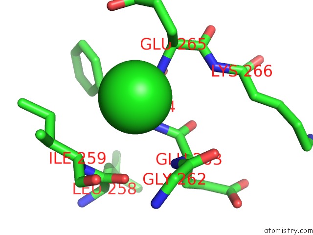
Mono view
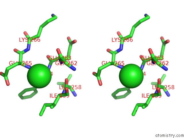
Stereo pair view
Mono view
Stereo pair view
A full contact list of Chlorine with other atoms in the Cl binding
site number 1 of Crystal Structure of A Meab and RV1496 Ortholog From Mycobacterium Thermoresistible Bound to Gdp within 5.0Å range:
|
Reference:
T.E.Edwards,
L.Baugh,
J.Bullen,
R.O.Baydo,
P.Witte,
K.Thompkins,
I.Q.Phan,
J.Abendroth,
M.C.Clifton,
B.Sankaran,
W.C.Van Voorhis,
P.J.Myler,
B.L.Staker,
C.Grundner,
D.D.Lorimer.
Crystal Structures of Mycobacterial Meab and Mmaa-Like Gtpases. J.Struct.Funct.Genom. V. 16 91 2015.
ISSN: ISSN 1345-711X
PubMed: 25832174
DOI: 10.1007/S10969-015-9197-2
Page generated: Sun Jul 21 05:17:39 2024
ISSN: ISSN 1345-711X
PubMed: 25832174
DOI: 10.1007/S10969-015-9197-2
Last articles
Zn in 9J0NZn in 9J0O
Zn in 9J0P
Zn in 9FJX
Zn in 9EKB
Zn in 9C0F
Zn in 9CAH
Zn in 9CH0
Zn in 9CH3
Zn in 9CH1