Chlorine »
PDB 3th4-3tnn »
3tkd »
Chlorine in PDB 3tkd: Crystal Structure of the GLUA2 Ligand-Binding Domain (S1S2J-L483Y- N754S) in Complex with Glutamate and Cyclothiazide at 1.45 A Resolution
Protein crystallography data
The structure of Crystal Structure of the GLUA2 Ligand-Binding Domain (S1S2J-L483Y- N754S) in Complex with Glutamate and Cyclothiazide at 1.45 A Resolution, PDB code: 3tkd
was solved by
C.Krintel,
K.Frydenvang,
M.Gajhede,
J.S.Kastrup,
with X-Ray Crystallography technique. A brief refinement statistics is given in the table below:
| Resolution Low / High (Å) | 26.26 / 1.45 |
| Space group | P 21 21 2 |
| Cell size a, b, c (Å), α, β, γ (°) | 98.197, 120.865, 47.232, 90.00, 90.00, 90.00 |
| R / Rfree (%) | 14.6 / 18 |
Chlorine Binding Sites:
The binding sites of Chlorine atom in the Crystal Structure of the GLUA2 Ligand-Binding Domain (S1S2J-L483Y- N754S) in Complex with Glutamate and Cyclothiazide at 1.45 A Resolution
(pdb code 3tkd). This binding sites where shown within
5.0 Angstroms radius around Chlorine atom.
In total 2 binding sites of Chlorine where determined in the Crystal Structure of the GLUA2 Ligand-Binding Domain (S1S2J-L483Y- N754S) in Complex with Glutamate and Cyclothiazide at 1.45 A Resolution, PDB code: 3tkd:
Jump to Chlorine binding site number: 1; 2;
In total 2 binding sites of Chlorine where determined in the Crystal Structure of the GLUA2 Ligand-Binding Domain (S1S2J-L483Y- N754S) in Complex with Glutamate and Cyclothiazide at 1.45 A Resolution, PDB code: 3tkd:
Jump to Chlorine binding site number: 1; 2;
Chlorine binding site 1 out of 2 in 3tkd
Go back to
Chlorine binding site 1 out
of 2 in the Crystal Structure of the GLUA2 Ligand-Binding Domain (S1S2J-L483Y- N754S) in Complex with Glutamate and Cyclothiazide at 1.45 A Resolution
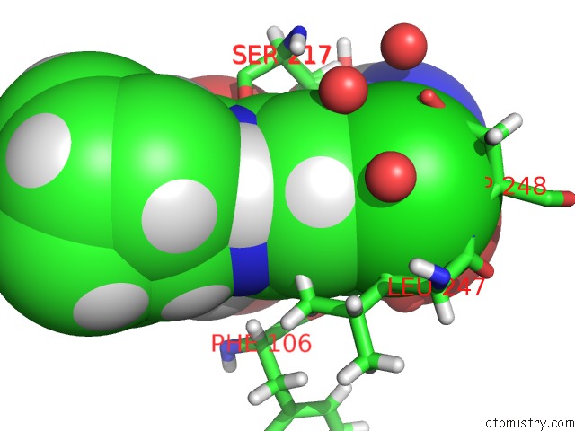
Mono view
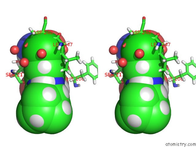
Stereo pair view
Mono view
Stereo pair view
A full contact list of Chlorine with other atoms in the Cl binding
site number 1 of Crystal Structure of the GLUA2 Ligand-Binding Domain (S1S2J-L483Y- N754S) in Complex with Glutamate and Cyclothiazide at 1.45 A Resolution within 5.0Å range:
|
Chlorine binding site 2 out of 2 in 3tkd
Go back to
Chlorine binding site 2 out
of 2 in the Crystal Structure of the GLUA2 Ligand-Binding Domain (S1S2J-L483Y- N754S) in Complex with Glutamate and Cyclothiazide at 1.45 A Resolution
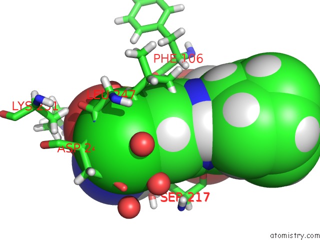
Mono view
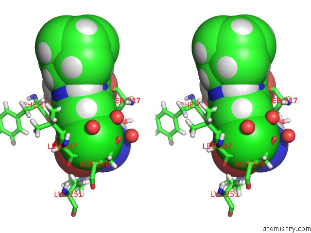
Stereo pair view
Mono view
Stereo pair view
A full contact list of Chlorine with other atoms in the Cl binding
site number 2 of Crystal Structure of the GLUA2 Ligand-Binding Domain (S1S2J-L483Y- N754S) in Complex with Glutamate and Cyclothiazide at 1.45 A Resolution within 5.0Å range:
|
Reference:
C.Krintel,
K.Frydenvang,
L.Olsen,
M.T.Kristensen,
O.De Barrios,
P.Naur,
P.Francotte,
B.Pirotte,
M.Gajhede,
J.S.Kastrup.
Thermodynamics and Structural Analysis of Positive Allosteric Modulation of the Ionotropic Glutamate Receptor GLUA2. Biochem.J. V. 441 173 2012.
ISSN: ISSN 0264-6021
PubMed: 21895609
DOI: 10.1042/BJ20111221
Page generated: Sun Jul 21 05:19:24 2024
ISSN: ISSN 0264-6021
PubMed: 21895609
DOI: 10.1042/BJ20111221
Last articles
Zn in 9J0NZn in 9J0O
Zn in 9J0P
Zn in 9FJX
Zn in 9EKB
Zn in 9C0F
Zn in 9CAH
Zn in 9CH0
Zn in 9CH3
Zn in 9CH1