Chlorine »
PDB 3txg-3u69 »
3tyk »
Chlorine in PDB 3tyk: Crystal Structure of Aminoglycoside Phosphotransferase Aph(4)-Ia
Enzymatic activity of Crystal Structure of Aminoglycoside Phosphotransferase Aph(4)-Ia
All present enzymatic activity of Crystal Structure of Aminoglycoside Phosphotransferase Aph(4)-Ia:
2.7.1.163;
2.7.1.163;
Protein crystallography data
The structure of Crystal Structure of Aminoglycoside Phosphotransferase Aph(4)-Ia, PDB code: 3tyk
was solved by
P.J.Stogios,
I.G.Shabalin,
T.Shakya,
E.Evdokmova,
Y.Fan,
M.Chruszcz,
W.Minor,
G.D.Wright,
A.Savchenko,
W.F.Anderson,
Midwest Center Forstructural Genomics (Mcsg),
with X-Ray Crystallography technique. A brief refinement statistics is given in the table below:
| Resolution Low / High (Å) | 34.01 / 1.95 |
| Space group | P 32 2 1 |
| Cell size a, b, c (Å), α, β, γ (°) | 70.640, 70.640, 125.880, 90.00, 90.00, 120.00 |
| R / Rfree (%) | 15.7 / 20.3 |
Chlorine Binding Sites:
The binding sites of Chlorine atom in the Crystal Structure of Aminoglycoside Phosphotransferase Aph(4)-Ia
(pdb code 3tyk). This binding sites where shown within
5.0 Angstroms radius around Chlorine atom.
In total only one binding site of Chlorine was determined in the Crystal Structure of Aminoglycoside Phosphotransferase Aph(4)-Ia, PDB code: 3tyk:
In total only one binding site of Chlorine was determined in the Crystal Structure of Aminoglycoside Phosphotransferase Aph(4)-Ia, PDB code: 3tyk:
Chlorine binding site 1 out of 1 in 3tyk
Go back to
Chlorine binding site 1 out
of 1 in the Crystal Structure of Aminoglycoside Phosphotransferase Aph(4)-Ia
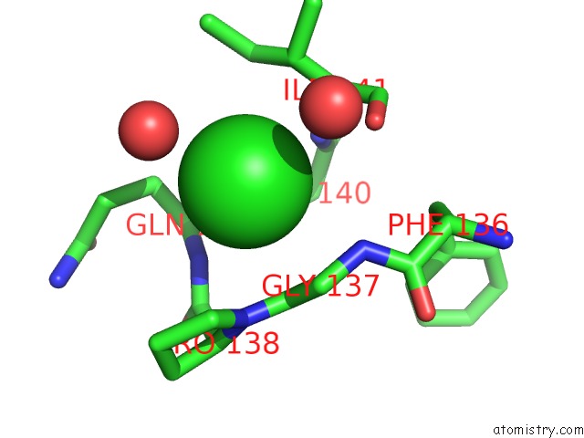
Mono view
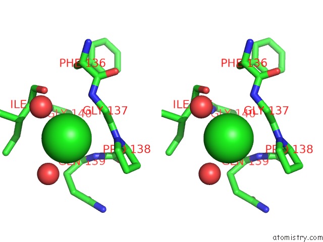
Stereo pair view
Mono view
Stereo pair view
A full contact list of Chlorine with other atoms in the Cl binding
site number 1 of Crystal Structure of Aminoglycoside Phosphotransferase Aph(4)-Ia within 5.0Å range:
|
Reference:
P.J.Stogios,
T.Shakya,
E.Evdokimova,
A.Savchenko,
G.D.Wright.
Structure and Function of Aph(4)-Ia, A Hygromycin B Resistance Enzyme. J.Biol.Chem. V. 286 1966 2011.
ISSN: ISSN 0021-9258
PubMed: 21084294
Page generated: Sun Jul 21 05:42:26 2024
ISSN: ISSN 0021-9258
PubMed: 21084294
Last articles
Zn in 9J0NZn in 9J0O
Zn in 9J0P
Zn in 9FJX
Zn in 9EKB
Zn in 9C0F
Zn in 9CAH
Zn in 9CH0
Zn in 9CH3
Zn in 9CH1