Chlorine »
PDB 3ue8-3unh »
3uha »
Chlorine in PDB 3uha: Crystal Structure of Saccharopine Dehydrogenase From Saccharomyces Cervisiae Complexed with Nad.
Enzymatic activity of Crystal Structure of Saccharopine Dehydrogenase From Saccharomyces Cervisiae Complexed with Nad.
All present enzymatic activity of Crystal Structure of Saccharopine Dehydrogenase From Saccharomyces Cervisiae Complexed with Nad.:
1.5.1.7;
1.5.1.7;
Protein crystallography data
The structure of Crystal Structure of Saccharopine Dehydrogenase From Saccharomyces Cervisiae Complexed with Nad., PDB code: 3uha
was solved by
P.F.Cook,
V.P.Kumar,
L.M.Thomas,
A.H.West,
K.D.Bobyk,
with X-Ray Crystallography technique. A brief refinement statistics is given in the table below:
| Resolution Low / High (Å) | 32.64 / 2.30 |
| Space group | P 1 21 1 |
| Cell size a, b, c (Å), α, β, γ (°) | 64.010, 104.450, 69.080, 90.00, 116.60, 90.00 |
| R / Rfree (%) | 20.7 / 27 |
Chlorine Binding Sites:
The binding sites of Chlorine atom in the Crystal Structure of Saccharopine Dehydrogenase From Saccharomyces Cervisiae Complexed with Nad.
(pdb code 3uha). This binding sites where shown within
5.0 Angstroms radius around Chlorine atom.
In total only one binding site of Chlorine was determined in the Crystal Structure of Saccharopine Dehydrogenase From Saccharomyces Cervisiae Complexed with Nad., PDB code: 3uha:
In total only one binding site of Chlorine was determined in the Crystal Structure of Saccharopine Dehydrogenase From Saccharomyces Cervisiae Complexed with Nad., PDB code: 3uha:
Chlorine binding site 1 out of 1 in 3uha
Go back to
Chlorine binding site 1 out
of 1 in the Crystal Structure of Saccharopine Dehydrogenase From Saccharomyces Cervisiae Complexed with Nad.
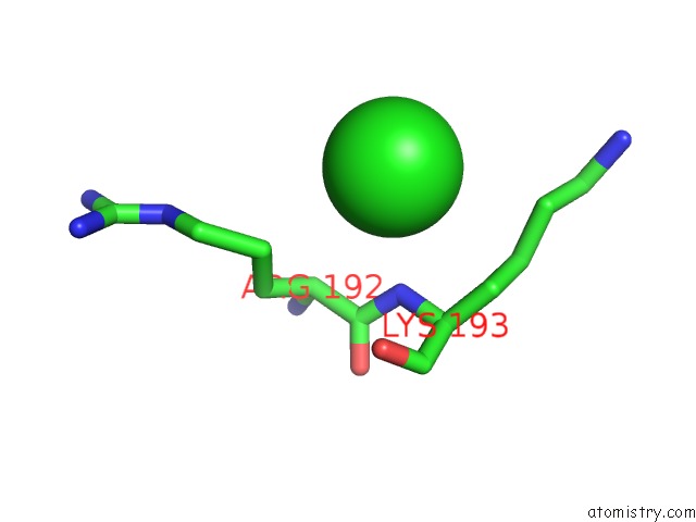
Mono view
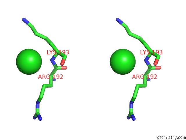
Stereo pair view
Mono view
Stereo pair view
A full contact list of Chlorine with other atoms in the Cl binding
site number 1 of Crystal Structure of Saccharopine Dehydrogenase From Saccharomyces Cervisiae Complexed with Nad. within 5.0Å range:
|
Reference:
V.P.Kumar,
L.M.Thomas,
K.D.Bobyk,
B.Andi,
P.F.Cook,
A.H.West.
Evidence in Support of Lysine 77 and Histidine 96 As Acid-Base Catalytic Residues in Saccharopine Dehydrogenase From Saccharomyces Cerevisiae. Biochemistry V. 51 857 2012.
ISSN: ISSN 0006-2960
PubMed: 22243403
DOI: 10.1021/BI201808U
Page generated: Sun Jul 21 06:07:17 2024
ISSN: ISSN 0006-2960
PubMed: 22243403
DOI: 10.1021/BI201808U
Last articles
Zn in 9J0NZn in 9J0O
Zn in 9J0P
Zn in 9FJX
Zn in 9EKB
Zn in 9C0F
Zn in 9CAH
Zn in 9CH0
Zn in 9CH3
Zn in 9CH1