Chlorine »
PDB 3v6e-3vfb »
3vav »
Chlorine in PDB 3vav: Crystal Structure of 3-Methyl-2-Oxobutanoate Hydroxymethyltransferase From Burkholderia Thailandensis
Enzymatic activity of Crystal Structure of 3-Methyl-2-Oxobutanoate Hydroxymethyltransferase From Burkholderia Thailandensis
All present enzymatic activity of Crystal Structure of 3-Methyl-2-Oxobutanoate Hydroxymethyltransferase From Burkholderia Thailandensis:
2.1.2.11;
2.1.2.11;
Protein crystallography data
The structure of Crystal Structure of 3-Methyl-2-Oxobutanoate Hydroxymethyltransferase From Burkholderia Thailandensis, PDB code: 3vav
was solved by
Seattle Structural Genomics Center For Infectious Disease (Ssgcid),
with X-Ray Crystallography technique. A brief refinement statistics is given in the table below:
| Resolution Low / High (Å) | 50.00 / 1.80 |
| Space group | P 1 21 1 |
| Cell size a, b, c (Å), α, β, γ (°) | 81.710, 174.750, 104.580, 90.00, 108.71, 90.00 |
| R / Rfree (%) | 15.4 / 18 |
Chlorine Binding Sites:
The binding sites of Chlorine atom in the Crystal Structure of 3-Methyl-2-Oxobutanoate Hydroxymethyltransferase From Burkholderia Thailandensis
(pdb code 3vav). This binding sites where shown within
5.0 Angstroms radius around Chlorine atom.
In total only one binding site of Chlorine was determined in the Crystal Structure of 3-Methyl-2-Oxobutanoate Hydroxymethyltransferase From Burkholderia Thailandensis, PDB code: 3vav:
In total only one binding site of Chlorine was determined in the Crystal Structure of 3-Methyl-2-Oxobutanoate Hydroxymethyltransferase From Burkholderia Thailandensis, PDB code: 3vav:
Chlorine binding site 1 out of 1 in 3vav
Go back to
Chlorine binding site 1 out
of 1 in the Crystal Structure of 3-Methyl-2-Oxobutanoate Hydroxymethyltransferase From Burkholderia Thailandensis
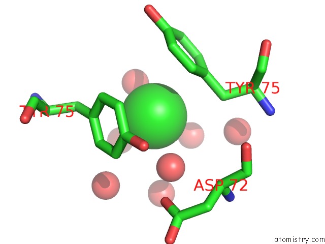
Mono view
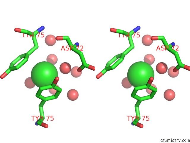
Stereo pair view
Mono view
Stereo pair view
A full contact list of Chlorine with other atoms in the Cl binding
site number 1 of Crystal Structure of 3-Methyl-2-Oxobutanoate Hydroxymethyltransferase From Burkholderia Thailandensis within 5.0Å range:
|
Reference:
L.Baugh,
L.A.Gallagher,
R.Patrapuvich,
M.C.Clifton,
A.S.Gardberg,
T.E.Edwards,
B.Armour,
D.W.Begley,
S.H.Dieterich,
D.M.Dranow,
J.Abendroth,
J.W.Fairman,
D.Fox,
B.L.Staker,
I.Phan,
A.Gillespie,
R.Choi,
S.Nakazawa-Hewitt,
M.T.Nguyen,
A.Napuli,
L.Barrett,
G.W.Buchko,
R.Stacy,
P.J.Myler,
L.J.Stewart,
C.Manoil,
W.C.Van Voorhis.
Combining Functional and Structural Genomics to Sample the Essential Burkholderia Structome. Plos One V. 8 53851 2013.
ISSN: ESSN 1932-6203
PubMed: 23382856
DOI: 10.1371/JOURNAL.PONE.0053851
Page generated: Sun Jul 21 06:47:20 2024
ISSN: ESSN 1932-6203
PubMed: 23382856
DOI: 10.1371/JOURNAL.PONE.0053851
Last articles
Ca in 5S5XCa in 5S5Y
Ca in 5S5W
Ca in 5S5V
Ca in 5S5U
Ca in 5S5T
Ca in 5S5S
Ca in 5S5R
Ca in 5S5Q
Ca in 5S5P