Chlorine »
PDB 3v6e-3vfb »
3vdh »
Chlorine in PDB 3vdh: Crystal Structure of PBGH5A, A Glycoside Hydrolase Family 5 Enzyme From Prevotella Bryantii B14
Protein crystallography data
The structure of Crystal Structure of PBGH5A, A Glycoside Hydrolase Family 5 Enzyme From Prevotella Bryantii B14, PDB code: 3vdh
was solved by
P.J.Stogios,
E.Evdokimova,
O.Egorova,
V.Yim,
A.Joachimiak,
A.M.Edwards,
A.Savchenko,
Midwest Center For Structural Genomics (Mcsg),
with X-Ray Crystallography technique. A brief refinement statistics is given in the table below:
| Resolution Low / High (Å) | 26.35 / 1.62 |
| Space group | P 1 21 1 |
| Cell size a, b, c (Å), α, β, γ (°) | 57.908, 85.254, 74.556, 90.00, 109.15, 90.00 |
| R / Rfree (%) | 15.1 / 19.9 |
Chlorine Binding Sites:
The binding sites of Chlorine atom in the Crystal Structure of PBGH5A, A Glycoside Hydrolase Family 5 Enzyme From Prevotella Bryantii B14
(pdb code 3vdh). This binding sites where shown within
5.0 Angstroms radius around Chlorine atom.
In total 3 binding sites of Chlorine where determined in the Crystal Structure of PBGH5A, A Glycoside Hydrolase Family 5 Enzyme From Prevotella Bryantii B14, PDB code: 3vdh:
Jump to Chlorine binding site number: 1; 2; 3;
In total 3 binding sites of Chlorine where determined in the Crystal Structure of PBGH5A, A Glycoside Hydrolase Family 5 Enzyme From Prevotella Bryantii B14, PDB code: 3vdh:
Jump to Chlorine binding site number: 1; 2; 3;
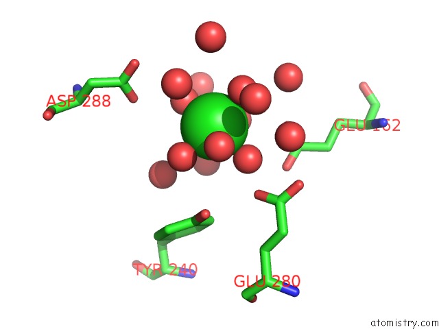
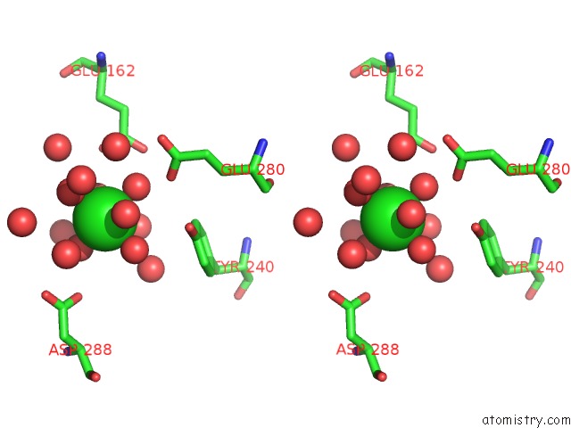
Chlorine binding site 1 out of 3 in 3vdh
Go back to
Chlorine binding site 1 out
of 3 in the Crystal Structure of PBGH5A, A Glycoside Hydrolase Family 5 Enzyme From Prevotella Bryantii B14
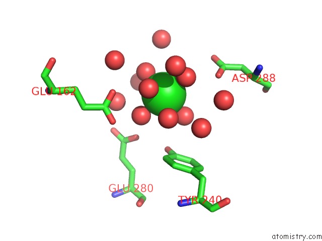
Mono view
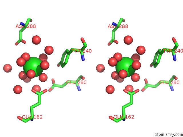
Stereo pair view
Mono view
Stereo pair view
A full contact list of Chlorine with other atoms in the Cl binding
site number 1 of Crystal Structure of PBGH5A, A Glycoside Hydrolase Family 5 Enzyme From Prevotella Bryantii B14 within 5.0Å range:
|
Chlorine binding site 2 out of 3 in 3vdh
Go back to
Chlorine binding site 2 out
of 3 in the Crystal Structure of PBGH5A, A Glycoside Hydrolase Family 5 Enzyme From Prevotella Bryantii B14
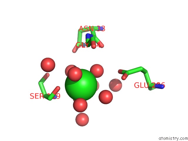
Mono view
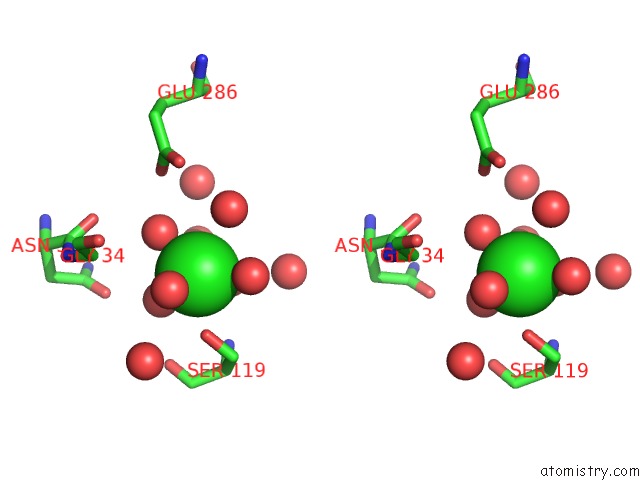
Stereo pair view
Mono view
Stereo pair view
A full contact list of Chlorine with other atoms in the Cl binding
site number 2 of Crystal Structure of PBGH5A, A Glycoside Hydrolase Family 5 Enzyme From Prevotella Bryantii B14 within 5.0Å range:
|
Chlorine binding site 3 out of 3 in 3vdh
Go back to
Chlorine binding site 3 out
of 3 in the Crystal Structure of PBGH5A, A Glycoside Hydrolase Family 5 Enzyme From Prevotella Bryantii B14
Mono view
Stereo pair view
Mono view
Stereo pair view
A full contact list of Chlorine with other atoms in the Cl binding
site number 3 of Crystal Structure of PBGH5A, A Glycoside Hydrolase Family 5 Enzyme From Prevotella Bryantii B14 within 5.0Å range:
|
Reference:
N.Mcgregor,
M.Morar,
T.H.Fenger,
P.Stogios,
N.Lenfant,
V.Yin,
X.Xu,
E.Evdokimova,
H.Cui,
B.Henrissat,
A.Savchenko,
H.Brumer.
Structure-Function Analysis of A Mixed-Linkage Beta-Glucanase/Xyloglucanase From the Key Ruminal Bacteroidetes Prevotella Bryantii B14. J.Biol.Chem. V. 291 1175 2016.
ISSN: ISSN 0021-9258
PubMed: 26507654
DOI: 10.1074/JBC.M115.691659
Page generated: Fri Jul 11 11:38:35 2025
ISSN: ISSN 0021-9258
PubMed: 26507654
DOI: 10.1074/JBC.M115.691659
Last articles
Cl in 4N4PCl in 4N78
Cl in 4N7S
Cl in 4N6X
Cl in 4N7I
Cl in 4N6P
Cl in 4N6D
Cl in 4N5H
Cl in 4N59
Cl in 4N2O