Chlorine »
PDB 3vfb-3vm1 »
3vik »
Chlorine in PDB 3vik: Crystal Structure of Beta-Glucosidase From Termite Neotermes Koshunensis in Complex with Cellobiose
Enzymatic activity of Crystal Structure of Beta-Glucosidase From Termite Neotermes Koshunensis in Complex with Cellobiose
All present enzymatic activity of Crystal Structure of Beta-Glucosidase From Termite Neotermes Koshunensis in Complex with Cellobiose:
3.2.1.21;
3.2.1.21;
Protein crystallography data
The structure of Crystal Structure of Beta-Glucosidase From Termite Neotermes Koshunensis in Complex with Cellobiose, PDB code: 3vik
was solved by
W.Y.Jeng,
C.I.Liu,
A.H.J.Wang,
with X-Ray Crystallography technique. A brief refinement statistics is given in the table below:
| Resolution Low / High (Å) | 22.95 / 1.10 |
| Space group | C 1 2 1 |
| Cell size a, b, c (Å), α, β, γ (°) | 92.965, 68.714, 75.832, 90.00, 95.73, 90.00 |
| R / Rfree (%) | 14.7 / 16.8 |
Other elements in 3vik:
The structure of Crystal Structure of Beta-Glucosidase From Termite Neotermes Koshunensis in Complex with Cellobiose also contains other interesting chemical elements:
| Sodium | (Na) | 1 atom |
Chlorine Binding Sites:
The binding sites of Chlorine atom in the Crystal Structure of Beta-Glucosidase From Termite Neotermes Koshunensis in Complex with Cellobiose
(pdb code 3vik). This binding sites where shown within
5.0 Angstroms radius around Chlorine atom.
In total only one binding site of Chlorine was determined in the Crystal Structure of Beta-Glucosidase From Termite Neotermes Koshunensis in Complex with Cellobiose, PDB code: 3vik:
In total only one binding site of Chlorine was determined in the Crystal Structure of Beta-Glucosidase From Termite Neotermes Koshunensis in Complex with Cellobiose, PDB code: 3vik:
Chlorine binding site 1 out of 1 in 3vik
Go back to
Chlorine binding site 1 out
of 1 in the Crystal Structure of Beta-Glucosidase From Termite Neotermes Koshunensis in Complex with Cellobiose
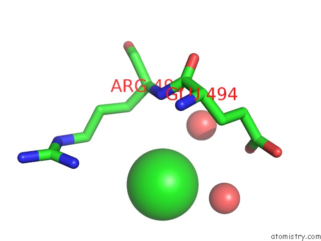
Mono view
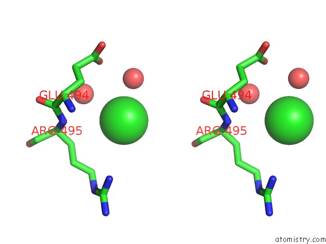
Stereo pair view
Mono view
Stereo pair view
A full contact list of Chlorine with other atoms in the Cl binding
site number 1 of Crystal Structure of Beta-Glucosidase From Termite Neotermes Koshunensis in Complex with Cellobiose within 5.0Å range:
|
Reference:
W.Y.Jeng,
N.C.Wang,
C.T.Lin,
W.J.Chang,
C.I.Liu,
A.H.J.Wang.
High-Resolution Structures of Neotermes Koshunensis Beta-Glucosidase Mutants Provide Insights Into the Catalytic Mechanism and the Synthesis of Glucoconjugates Acta Crystallogr.,Sect.D V. 68 829 2012.
ISSN: ISSN 0907-4449
PubMed: 22751668
DOI: 10.1107/S0907444912013224
Page generated: Sun Jul 21 06:58:15 2024
ISSN: ISSN 0907-4449
PubMed: 22751668
DOI: 10.1107/S0907444912013224
Last articles
Zn in 9JYWZn in 9IR4
Zn in 9IR3
Zn in 9GMX
Zn in 9GMW
Zn in 9JEJ
Zn in 9ERF
Zn in 9ERE
Zn in 9EGV
Zn in 9EGW