Chlorine »
PDB 3vm5-3vw8 »
3vm6 »
Chlorine in PDB 3vm6: Crystal Structure of Ribose-1,5-Bisphosphate Isomerase From Thermococcus Kodakarensis KOD1 in Complex with Alpha-D-Ribose-1,5- Bisphosphate
Protein crystallography data
The structure of Crystal Structure of Ribose-1,5-Bisphosphate Isomerase From Thermococcus Kodakarensis KOD1 in Complex with Alpha-D-Ribose-1,5- Bisphosphate, PDB code: 3vm6
was solved by
A.Nakamura,
M.Fujihashi,
R.Aono,
T.Sato,
Y.Nishiba,
S.Yoshida,
A.Yano,
H.Atomi,
T.Imanaka,
K.Miki,
with X-Ray Crystallography technique. A brief refinement statistics is given in the table below:
| Resolution Low / High (Å) | 45.97 / 2.85 |
| Space group | P 41 21 2 |
| Cell size a, b, c (Å), α, β, γ (°) | 146.537, 146.537, 99.649, 90.00, 90.00, 90.00 |
| R / Rfree (%) | 20.1 / 26.2 |
Other elements in 3vm6:
The structure of Crystal Structure of Ribose-1,5-Bisphosphate Isomerase From Thermococcus Kodakarensis KOD1 in Complex with Alpha-D-Ribose-1,5- Bisphosphate also contains other interesting chemical elements:
| Magnesium | (Mg) | 2 atoms |
Chlorine Binding Sites:
The binding sites of Chlorine atom in the Crystal Structure of Ribose-1,5-Bisphosphate Isomerase From Thermococcus Kodakarensis KOD1 in Complex with Alpha-D-Ribose-1,5- Bisphosphate
(pdb code 3vm6). This binding sites where shown within
5.0 Angstroms radius around Chlorine atom.
In total 3 binding sites of Chlorine where determined in the Crystal Structure of Ribose-1,5-Bisphosphate Isomerase From Thermococcus Kodakarensis KOD1 in Complex with Alpha-D-Ribose-1,5- Bisphosphate, PDB code: 3vm6:
Jump to Chlorine binding site number: 1; 2; 3;
In total 3 binding sites of Chlorine where determined in the Crystal Structure of Ribose-1,5-Bisphosphate Isomerase From Thermococcus Kodakarensis KOD1 in Complex with Alpha-D-Ribose-1,5- Bisphosphate, PDB code: 3vm6:
Jump to Chlorine binding site number: 1; 2; 3;
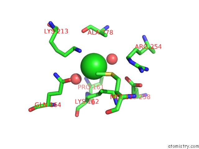
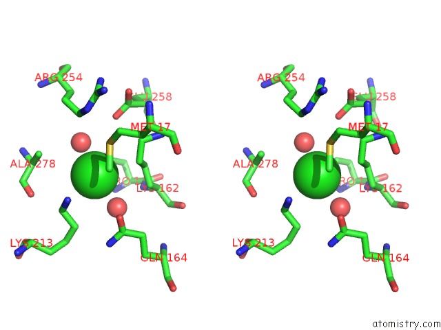
Chlorine binding site 1 out of 3 in 3vm6
Go back to
Chlorine binding site 1 out
of 3 in the Crystal Structure of Ribose-1,5-Bisphosphate Isomerase From Thermococcus Kodakarensis KOD1 in Complex with Alpha-D-Ribose-1,5- Bisphosphate
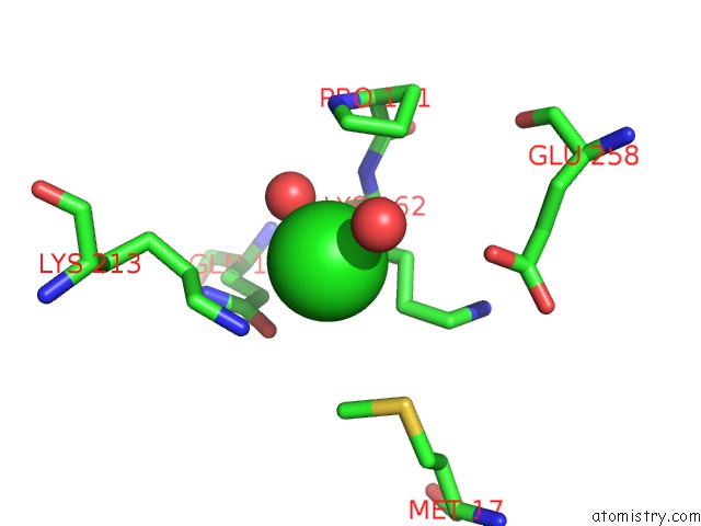
Mono view
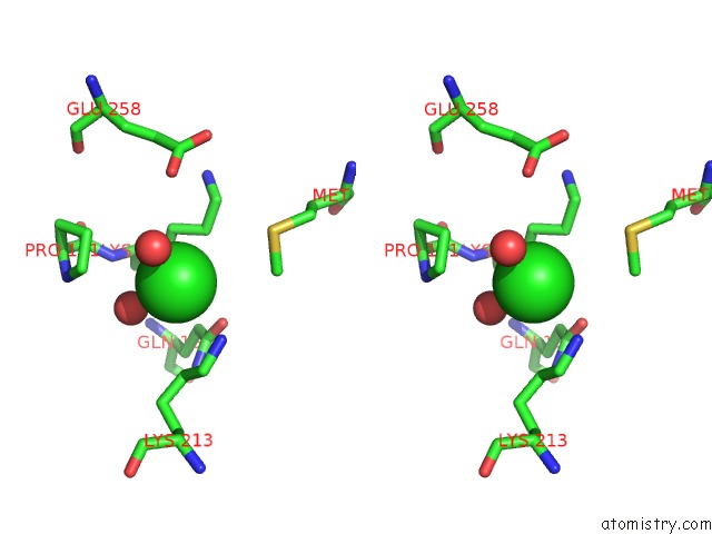
Stereo pair view
Mono view
Stereo pair view
A full contact list of Chlorine with other atoms in the Cl binding
site number 1 of Crystal Structure of Ribose-1,5-Bisphosphate Isomerase From Thermococcus Kodakarensis KOD1 in Complex with Alpha-D-Ribose-1,5- Bisphosphate within 5.0Å range:
|
Chlorine binding site 2 out of 3 in 3vm6
Go back to
Chlorine binding site 2 out
of 3 in the Crystal Structure of Ribose-1,5-Bisphosphate Isomerase From Thermococcus Kodakarensis KOD1 in Complex with Alpha-D-Ribose-1,5- Bisphosphate
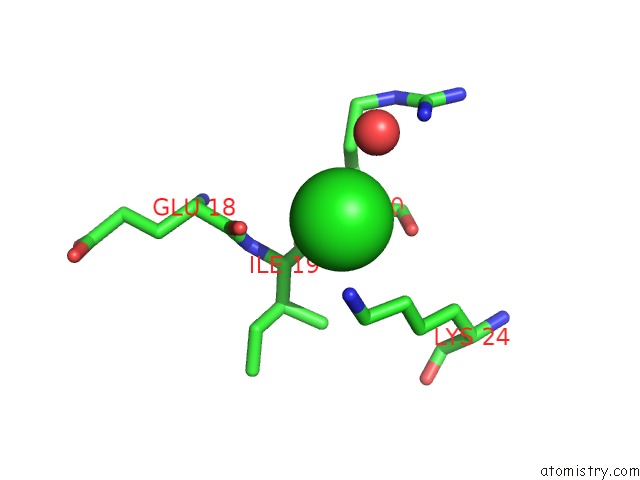
Mono view
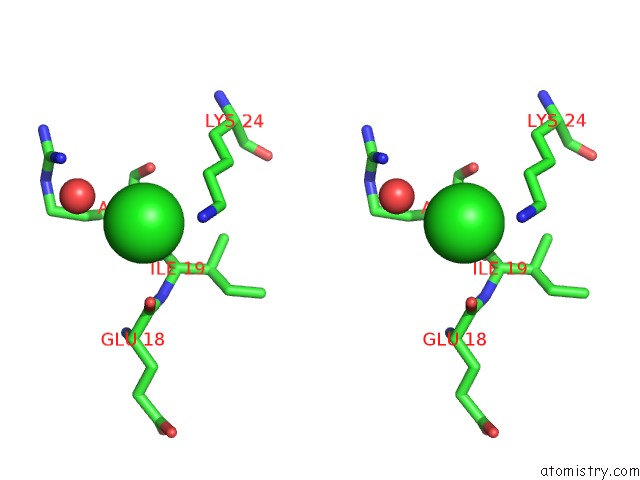
Stereo pair view
Mono view
Stereo pair view
A full contact list of Chlorine with other atoms in the Cl binding
site number 2 of Crystal Structure of Ribose-1,5-Bisphosphate Isomerase From Thermococcus Kodakarensis KOD1 in Complex with Alpha-D-Ribose-1,5- Bisphosphate within 5.0Å range:
|
Chlorine binding site 3 out of 3 in 3vm6
Go back to
Chlorine binding site 3 out
of 3 in the Crystal Structure of Ribose-1,5-Bisphosphate Isomerase From Thermococcus Kodakarensis KOD1 in Complex with Alpha-D-Ribose-1,5- Bisphosphate
Mono view
Stereo pair view
Mono view
Stereo pair view
A full contact list of Chlorine with other atoms in the Cl binding
site number 3 of Crystal Structure of Ribose-1,5-Bisphosphate Isomerase From Thermococcus Kodakarensis KOD1 in Complex with Alpha-D-Ribose-1,5- Bisphosphate within 5.0Å range:
|
Reference:
A.Nakamura,
M.Fujihashi,
R.Aono,
T.Sato,
Y.Nishiba,
S.Yoshida,
A.Yano,
H.Atomi,
T.Imanaka,
K.Miki.
Dynamic, Ligand-Dependent Conformational Change Triggers Reaction of Ribose-1,5-Bisphosphate Isomerase From Thermococcus Kodakarensis KOD1 J.Biol.Chem. V. 287 20784 2012.
ISSN: ISSN 0021-9258
PubMed: 22511789
DOI: 10.1074/JBC.M112.349423
Page generated: Sun Jul 21 07:05:50 2024
ISSN: ISSN 0021-9258
PubMed: 22511789
DOI: 10.1074/JBC.M112.349423
Last articles
Zn in 9JYWZn in 9IR4
Zn in 9IR3
Zn in 9GMX
Zn in 9GMW
Zn in 9JEJ
Zn in 9ERF
Zn in 9ERE
Zn in 9EGV
Zn in 9EGW