Chlorine »
PDB 3vm6-3vwd »
3vot »
Chlorine in PDB 3vot: Crystal Structure of L-Amino Acid Ligase From Bacillus Licheniformis
Enzymatic activity of Crystal Structure of L-Amino Acid Ligase From Bacillus Licheniformis
All present enzymatic activity of Crystal Structure of L-Amino Acid Ligase From Bacillus Licheniformis:
6.3.2.28;
6.3.2.28;
Protein crystallography data
The structure of Crystal Structure of L-Amino Acid Ligase From Bacillus Licheniformis, PDB code: 3vot
was solved by
M.Suzuki,
Y.Takahashi,
A.Noguchi,
T.Arai,
M.Yagasaki,
K.Kino,
J.Saito,
with X-Ray Crystallography technique. A brief refinement statistics is given in the table below:
| Resolution Low / High (Å) | 29.46 / 1.80 |
| Space group | P 21 21 21 |
| Cell size a, b, c (Å), α, β, γ (°) | 58.090, 90.030, 154.910, 90.00, 90.00, 90.00 |
| R / Rfree (%) | 19.6 / 24.1 |
Other elements in 3vot:
The structure of Crystal Structure of L-Amino Acid Ligase From Bacillus Licheniformis also contains other interesting chemical elements:
| Calcium | (Ca) | 9 atoms |
Chlorine Binding Sites:
The binding sites of Chlorine atom in the Crystal Structure of L-Amino Acid Ligase From Bacillus Licheniformis
(pdb code 3vot). This binding sites where shown within
5.0 Angstroms radius around Chlorine atom.
In total 2 binding sites of Chlorine where determined in the Crystal Structure of L-Amino Acid Ligase From Bacillus Licheniformis, PDB code: 3vot:
Jump to Chlorine binding site number: 1; 2;
In total 2 binding sites of Chlorine where determined in the Crystal Structure of L-Amino Acid Ligase From Bacillus Licheniformis, PDB code: 3vot:
Jump to Chlorine binding site number: 1; 2;
Chlorine binding site 1 out of 2 in 3vot
Go back to
Chlorine binding site 1 out
of 2 in the Crystal Structure of L-Amino Acid Ligase From Bacillus Licheniformis
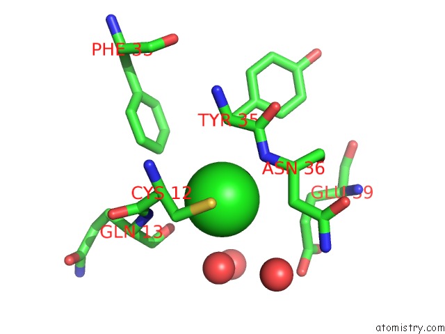
Mono view
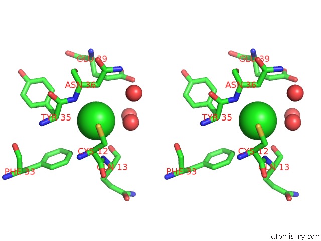
Stereo pair view
Mono view
Stereo pair view
A full contact list of Chlorine with other atoms in the Cl binding
site number 1 of Crystal Structure of L-Amino Acid Ligase From Bacillus Licheniformis within 5.0Å range:
|
Chlorine binding site 2 out of 2 in 3vot
Go back to
Chlorine binding site 2 out
of 2 in the Crystal Structure of L-Amino Acid Ligase From Bacillus Licheniformis
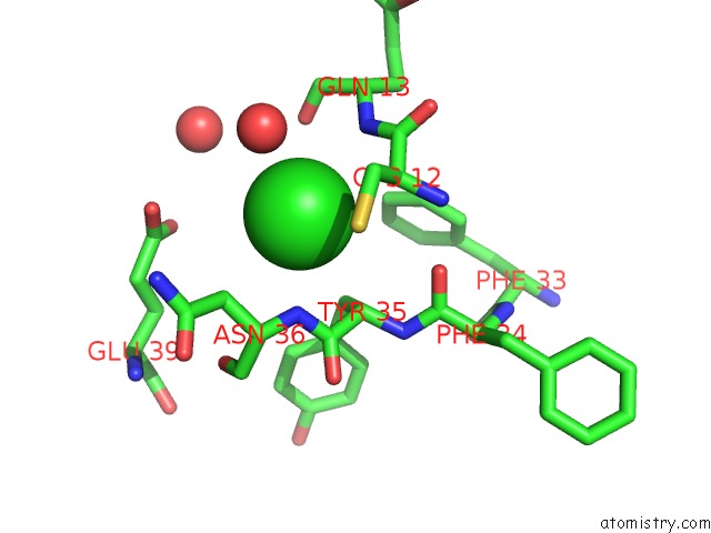
Mono view
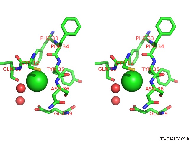
Stereo pair view
Mono view
Stereo pair view
A full contact list of Chlorine with other atoms in the Cl binding
site number 2 of Crystal Structure of L-Amino Acid Ligase From Bacillus Licheniformis within 5.0Å range:
|
Reference:
M.Suzuki,
Y.Takahashi,
A.Noguchi,
T.Arai,
M.Yagasaki,
K.Kino,
J.Saito.
The Structure of L-Amino-Acid Ligase From Bacillus Licheniformis Acta Crystallogr.,Sect.D V. 68 1535 2012.
ISSN: ISSN 0907-4449
PubMed: 23090402
DOI: 10.1107/S0907444912038103
Page generated: Sun Jul 21 07:07:24 2024
ISSN: ISSN 0907-4449
PubMed: 23090402
DOI: 10.1107/S0907444912038103
Last articles
Zn in 9J0NZn in 9J0O
Zn in 9J0P
Zn in 9FJX
Zn in 9EKB
Zn in 9C0F
Zn in 9CAH
Zn in 9CH0
Zn in 9CH3
Zn in 9CH1