Chlorine »
PDB 3vm6-3vwd »
3vu1 »
Chlorine in PDB 3vu1: Crystal Structure of the C-Terminal Globular Domain of Oligosaccharyltransferase (Phaglb-L, O74088_PYRHO) From Pyrococcus Horikoshii
Enzymatic activity of Crystal Structure of the C-Terminal Globular Domain of Oligosaccharyltransferase (Phaglb-L, O74088_PYRHO) From Pyrococcus Horikoshii
All present enzymatic activity of Crystal Structure of the C-Terminal Globular Domain of Oligosaccharyltransferase (Phaglb-L, O74088_PYRHO) From Pyrococcus Horikoshii:
2.4.1.119;
2.4.1.119;
Protein crystallography data
The structure of Crystal Structure of the C-Terminal Globular Domain of Oligosaccharyltransferase (Phaglb-L, O74088_PYRHO) From Pyrococcus Horikoshii, PDB code: 3vu1
was solved by
J.Nyirenda,
S.Matsumoto,
T.Saitoh,
N.Maita,
N.N.Noda,
F.Inagaki,
D.Kohda,
with X-Ray Crystallography technique. A brief refinement statistics is given in the table below:
| Resolution Low / High (Å) | 30.00 / 2.70 |
| Space group | P 21 21 21 |
| Cell size a, b, c (Å), α, β, γ (°) | 83.467, 94.844, 186.345, 90.00, 90.00, 90.00 |
| R / Rfree (%) | 17.1 / 21.5 |
Other elements in 3vu1:
The structure of Crystal Structure of the C-Terminal Globular Domain of Oligosaccharyltransferase (Phaglb-L, O74088_PYRHO) From Pyrococcus Horikoshii also contains other interesting chemical elements:
| Calcium | (Ca) | 2 atoms |
Chlorine Binding Sites:
The binding sites of Chlorine atom in the Crystal Structure of the C-Terminal Globular Domain of Oligosaccharyltransferase (Phaglb-L, O74088_PYRHO) From Pyrococcus Horikoshii
(pdb code 3vu1). This binding sites where shown within
5.0 Angstroms radius around Chlorine atom.
In total 2 binding sites of Chlorine where determined in the Crystal Structure of the C-Terminal Globular Domain of Oligosaccharyltransferase (Phaglb-L, O74088_PYRHO) From Pyrococcus Horikoshii, PDB code: 3vu1:
Jump to Chlorine binding site number: 1; 2;
In total 2 binding sites of Chlorine where determined in the Crystal Structure of the C-Terminal Globular Domain of Oligosaccharyltransferase (Phaglb-L, O74088_PYRHO) From Pyrococcus Horikoshii, PDB code: 3vu1:
Jump to Chlorine binding site number: 1; 2;
Chlorine binding site 1 out of 2 in 3vu1
Go back to
Chlorine binding site 1 out
of 2 in the Crystal Structure of the C-Terminal Globular Domain of Oligosaccharyltransferase (Phaglb-L, O74088_PYRHO) From Pyrococcus Horikoshii
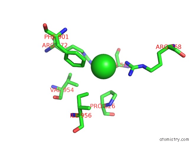
Mono view
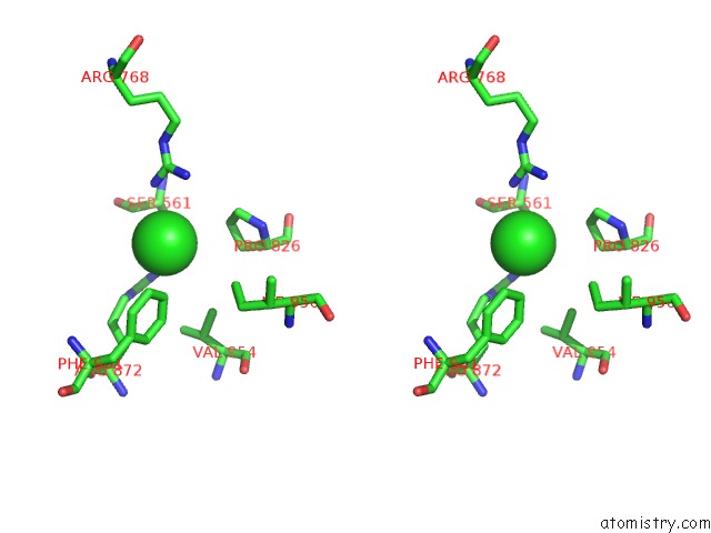
Stereo pair view
Mono view
Stereo pair view
A full contact list of Chlorine with other atoms in the Cl binding
site number 1 of Crystal Structure of the C-Terminal Globular Domain of Oligosaccharyltransferase (Phaglb-L, O74088_PYRHO) From Pyrococcus Horikoshii within 5.0Å range:
|
Chlorine binding site 2 out of 2 in 3vu1
Go back to
Chlorine binding site 2 out
of 2 in the Crystal Structure of the C-Terminal Globular Domain of Oligosaccharyltransferase (Phaglb-L, O74088_PYRHO) From Pyrococcus Horikoshii
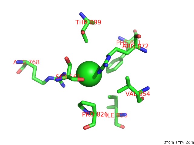
Mono view
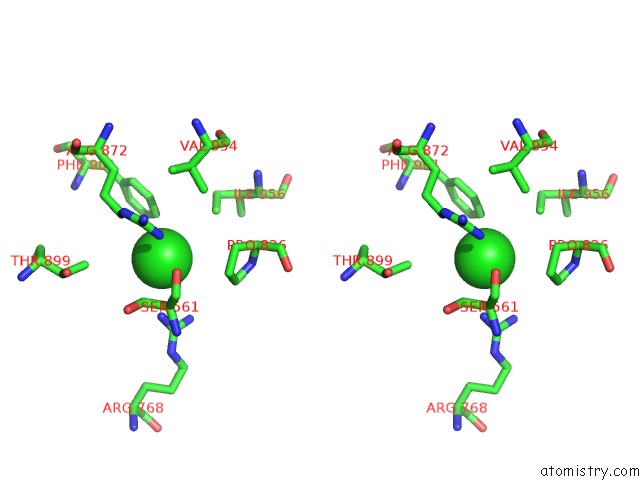
Stereo pair view
Mono view
Stereo pair view
A full contact list of Chlorine with other atoms in the Cl binding
site number 2 of Crystal Structure of the C-Terminal Globular Domain of Oligosaccharyltransferase (Phaglb-L, O74088_PYRHO) From Pyrococcus Horikoshii within 5.0Å range:
|
Reference:
J.Nyirenda,
S.Matsumoto,
T.Saitoh,
N.Maita,
N.N.Noda,
F.Inagaki,
D.Kohda.
Crystallographic and uc(Nmr) Evidence For Flexibility in Oligosaccharyltransferases and Its Catalytic Significance Structure V. 21 32 2013.
ISSN: ISSN 0969-2126
PubMed: 23177926
DOI: 10.1016/J.STR.2012.10.011
Page generated: Sun Jul 21 07:11:46 2024
ISSN: ISSN 0969-2126
PubMed: 23177926
DOI: 10.1016/J.STR.2012.10.011
Last articles
Zn in 9J0NZn in 9J0O
Zn in 9J0P
Zn in 9FJX
Zn in 9EKB
Zn in 9C0F
Zn in 9CAH
Zn in 9CH0
Zn in 9CH3
Zn in 9CH1