Chlorine »
PDB 3wav-3wnr »
3wn1 »
Chlorine in PDB 3wn1: Crystal Structure of Streptomyces Coelicolor Alpha-L- Arabinofuranosidase in Complex with Xylotriose
Enzymatic activity of Crystal Structure of Streptomyces Coelicolor Alpha-L- Arabinofuranosidase in Complex with Xylotriose
All present enzymatic activity of Crystal Structure of Streptomyces Coelicolor Alpha-L- Arabinofuranosidase in Complex with Xylotriose:
3.2.1.55;
3.2.1.55;
Protein crystallography data
The structure of Crystal Structure of Streptomyces Coelicolor Alpha-L- Arabinofuranosidase in Complex with Xylotriose, PDB code: 3wn1
was solved by
Z.Fujimoto,
T.Maehara,
H.Ichinose,
M.Michikawa,
K.Harazono,
S.Kaneko,
with X-Ray Crystallography technique. A brief refinement statistics is given in the table below:
| Resolution Low / High (Å) | 31.07 / 2.00 |
| Space group | P 41 21 2 |
| Cell size a, b, c (Å), α, β, γ (°) | 97.668, 97.668, 104.019, 90.00, 90.00, 90.00 |
| R / Rfree (%) | 17.9 / 20.4 |
Other elements in 3wn1:
The structure of Crystal Structure of Streptomyces Coelicolor Alpha-L- Arabinofuranosidase in Complex with Xylotriose also contains other interesting chemical elements:
| Calcium | (Ca) | 1 atom |
Chlorine Binding Sites:
The binding sites of Chlorine atom in the Crystal Structure of Streptomyces Coelicolor Alpha-L- Arabinofuranosidase in Complex with Xylotriose
(pdb code 3wn1). This binding sites where shown within
5.0 Angstroms radius around Chlorine atom.
In total only one binding site of Chlorine was determined in the Crystal Structure of Streptomyces Coelicolor Alpha-L- Arabinofuranosidase in Complex with Xylotriose, PDB code: 3wn1:
In total only one binding site of Chlorine was determined in the Crystal Structure of Streptomyces Coelicolor Alpha-L- Arabinofuranosidase in Complex with Xylotriose, PDB code: 3wn1:
Chlorine binding site 1 out of 1 in 3wn1
Go back to
Chlorine binding site 1 out
of 1 in the Crystal Structure of Streptomyces Coelicolor Alpha-L- Arabinofuranosidase in Complex with Xylotriose
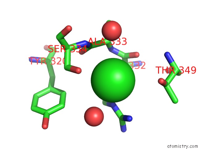
Mono view
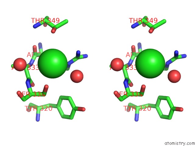
Stereo pair view
Mono view
Stereo pair view
A full contact list of Chlorine with other atoms in the Cl binding
site number 1 of Crystal Structure of Streptomyces Coelicolor Alpha-L- Arabinofuranosidase in Complex with Xylotriose within 5.0Å range:
|
Reference:
T.Maehara,
Z.Fujimoto,
H.Ichinose,
M.Michikawa,
K.Harazono,
S.Kaneko.
Crystal Structure and Characterization of the Glycoside Hydrolase Family 62 Alpha-L-Arabinofuranosidase From Streptomyces Coelicolor J.Biol.Chem. V. 289 7962 2014.
ISSN: ISSN 0021-9258
PubMed: 24482228
DOI: 10.1074/JBC.M113.540542
Page generated: Sun Jul 21 07:36:36 2024
ISSN: ISSN 0021-9258
PubMed: 24482228
DOI: 10.1074/JBC.M113.540542
Last articles
Zn in 9J0NZn in 9J0O
Zn in 9J0P
Zn in 9FJX
Zn in 9EKB
Zn in 9C0F
Zn in 9CAH
Zn in 9CH0
Zn in 9CH3
Zn in 9CH1