Chlorine »
PDB 3wns-3x1v »
3wtk »
Chlorine in PDB 3wtk: Crystal Structure of Lymnaea Stagnalis Acetylcholine-Binding Protein Q55R Mutant Complexed with Thiacloprid
Protein crystallography data
The structure of Crystal Structure of Lymnaea Stagnalis Acetylcholine-Binding Protein Q55R Mutant Complexed with Thiacloprid, PDB code: 3wtk
was solved by
T.Okajima,
M.Ihara,
A.Yamashita,
T.Oda,
K.Matsuda,
with X-Ray Crystallography technique. A brief refinement statistics is given in the table below:
| Resolution Low / High (Å) | 39.60 / 2.69 |
| Space group | P 65 |
| Cell size a, b, c (Å), α, β, γ (°) | 74.534, 74.534, 350.816, 90.00, 90.00, 120.00 |
| R / Rfree (%) | 20 / 27.4 |
Chlorine Binding Sites:
The binding sites of Chlorine atom in the Crystal Structure of Lymnaea Stagnalis Acetylcholine-Binding Protein Q55R Mutant Complexed with Thiacloprid
(pdb code 3wtk). This binding sites where shown within
5.0 Angstroms radius around Chlorine atom.
In total 5 binding sites of Chlorine where determined in the Crystal Structure of Lymnaea Stagnalis Acetylcholine-Binding Protein Q55R Mutant Complexed with Thiacloprid, PDB code: 3wtk:
Jump to Chlorine binding site number: 1; 2; 3; 4; 5;
In total 5 binding sites of Chlorine where determined in the Crystal Structure of Lymnaea Stagnalis Acetylcholine-Binding Protein Q55R Mutant Complexed with Thiacloprid, PDB code: 3wtk:
Jump to Chlorine binding site number: 1; 2; 3; 4; 5;
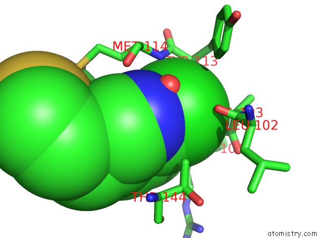
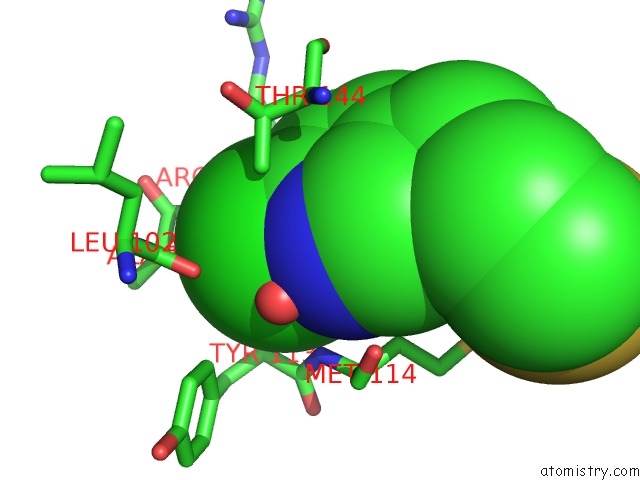
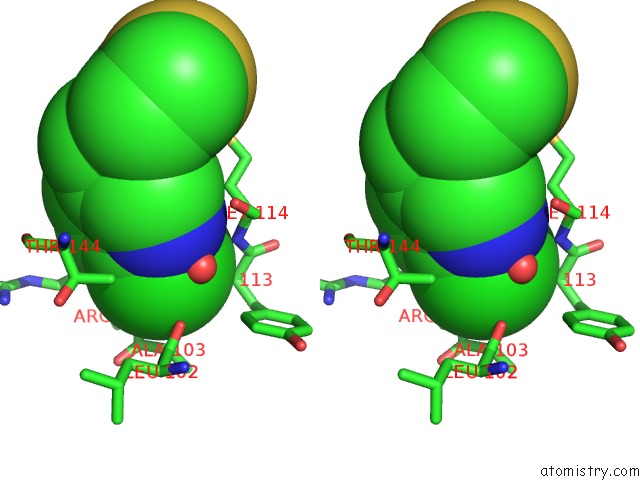
Chlorine binding site 1 out of 5 in 3wtk
Go back to
Chlorine binding site 1 out
of 5 in the Crystal Structure of Lymnaea Stagnalis Acetylcholine-Binding Protein Q55R Mutant Complexed with Thiacloprid
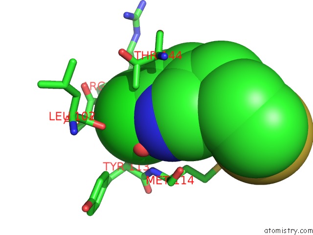
Mono view
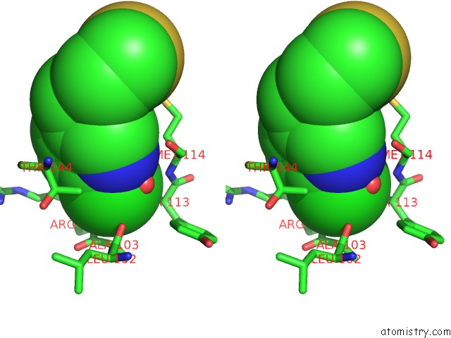
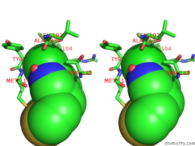
Stereo pair view
Mono view
Stereo pair view
A full contact list of Chlorine with other atoms in the Cl binding
site number 1 of Crystal Structure of Lymnaea Stagnalis Acetylcholine-Binding Protein Q55R Mutant Complexed with Thiacloprid within 5.0Å range:
|
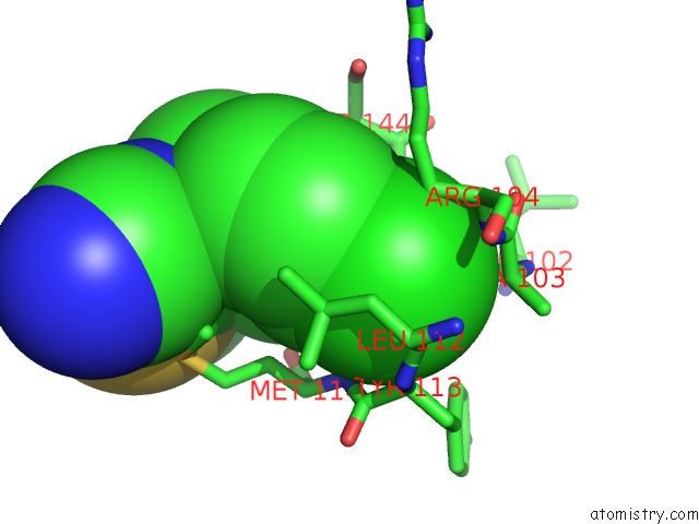
Chlorine binding site 2 out of 5 in 3wtk
Go back to
Chlorine binding site 2 out
of 5 in the Crystal Structure of Lymnaea Stagnalis Acetylcholine-Binding Protein Q55R Mutant Complexed with Thiacloprid
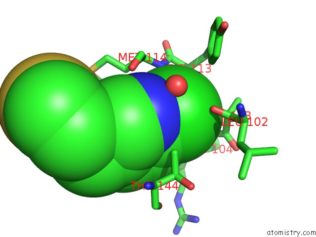
Mono view
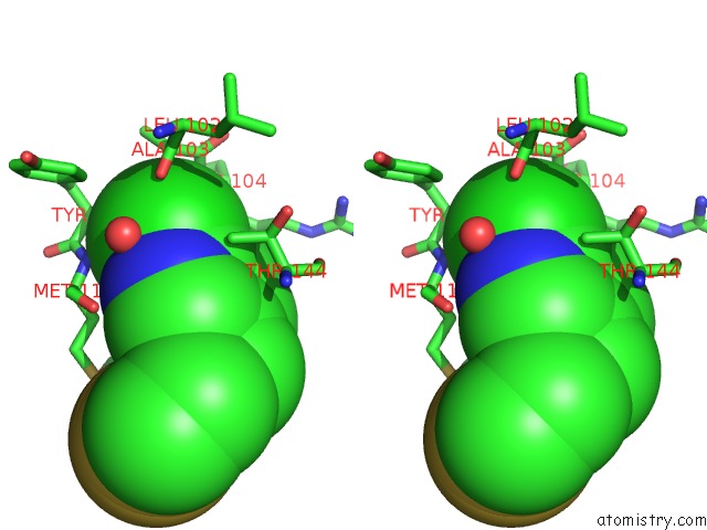
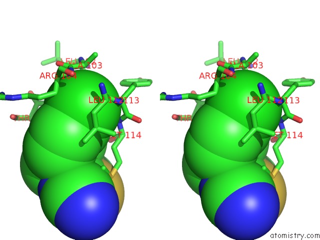
Stereo pair view
Mono view
Stereo pair view
A full contact list of Chlorine with other atoms in the Cl binding
site number 2 of Crystal Structure of Lymnaea Stagnalis Acetylcholine-Binding Protein Q55R Mutant Complexed with Thiacloprid within 5.0Å range:
|
Chlorine binding site 3 out of 5 in 3wtk
Go back to
Chlorine binding site 3 out
of 5 in the Crystal Structure of Lymnaea Stagnalis Acetylcholine-Binding Protein Q55R Mutant Complexed with Thiacloprid
Mono view
Stereo pair view
Mono view
Stereo pair view
A full contact list of Chlorine with other atoms in the Cl binding
site number 3 of Crystal Structure of Lymnaea Stagnalis Acetylcholine-Binding Protein Q55R Mutant Complexed with Thiacloprid within 5.0Å range:
|
Chlorine binding site 4 out of 5 in 3wtk
Go back to
Chlorine binding site 4 out
of 5 in the Crystal Structure of Lymnaea Stagnalis Acetylcholine-Binding Protein Q55R Mutant Complexed with Thiacloprid
Mono view
Stereo pair view
Mono view
Stereo pair view
A full contact list of Chlorine with other atoms in the Cl binding
site number 4 of Crystal Structure of Lymnaea Stagnalis Acetylcholine-Binding Protein Q55R Mutant Complexed with Thiacloprid within 5.0Å range:
|
Chlorine binding site 5 out of 5 in 3wtk
Go back to
Chlorine binding site 5 out
of 5 in the Crystal Structure of Lymnaea Stagnalis Acetylcholine-Binding Protein Q55R Mutant Complexed with Thiacloprid
Mono view
Stereo pair view
Mono view
Stereo pair view
A full contact list of Chlorine with other atoms in the Cl binding
site number 5 of Crystal Structure of Lymnaea Stagnalis Acetylcholine-Binding Protein Q55R Mutant Complexed with Thiacloprid within 5.0Å range:
|
Reference:
M.Ihara,
T.Okajima,
A.Yamashita,
T.Oda,
T.Asano,
M.Matsui,
D.B.Sattelle,
K.Matsuda.
Studies on An Acetylcholine Binding Protein Identify A Basic Residue in Loop G on the Beta 1 Strand As A New Structural Determinant of Neonicotinoid Actions Mol.Pharmacol. V. 86 736 2014.
ISSN: ISSN 0026-895X
PubMed: 25267717
DOI: 10.1124/MOL.114.094698
Page generated: Sun Jul 21 07:42:31 2024
ISSN: ISSN 0026-895X
PubMed: 25267717
DOI: 10.1124/MOL.114.094698
Last articles
Zn in 9J0NZn in 9J0O
Zn in 9J0P
Zn in 9FJX
Zn in 9EKB
Zn in 9C0F
Zn in 9CAH
Zn in 9CH0
Zn in 9CH3
Zn in 9CH1