Chlorine »
PDB 3wns-3x1v »
3ww6 »
Chlorine in PDB 3ww6: Crystal Structure of Hen Egg White Lysozyme Mutant N46D/D52S
Enzymatic activity of Crystal Structure of Hen Egg White Lysozyme Mutant N46D/D52S
All present enzymatic activity of Crystal Structure of Hen Egg White Lysozyme Mutant N46D/D52S:
3.2.1.17;
3.2.1.17;
Protein crystallography data
The structure of Crystal Structure of Hen Egg White Lysozyme Mutant N46D/D52S, PDB code: 3ww6
was solved by
Y.Abe,
M.Kubota,
Y.Ito,
T.Imoto,
T.Ueda,
with X-Ray Crystallography technique. A brief refinement statistics is given in the table below:
| Resolution Low / High (Å) | 26.75 / 1.53 |
| Space group | P 43 21 2 |
| Cell size a, b, c (Å), α, β, γ (°) | 77.006, 77.006, 37.185, 90.00, 90.00, 90.00 |
| R / Rfree (%) | 18.7 / 20.5 |
Chlorine Binding Sites:
The binding sites of Chlorine atom in the Crystal Structure of Hen Egg White Lysozyme Mutant N46D/D52S
(pdb code 3ww6). This binding sites where shown within
5.0 Angstroms radius around Chlorine atom.
In total 5 binding sites of Chlorine where determined in the Crystal Structure of Hen Egg White Lysozyme Mutant N46D/D52S, PDB code: 3ww6:
Jump to Chlorine binding site number: 1; 2; 3; 4; 5;
In total 5 binding sites of Chlorine where determined in the Crystal Structure of Hen Egg White Lysozyme Mutant N46D/D52S, PDB code: 3ww6:
Jump to Chlorine binding site number: 1; 2; 3; 4; 5;
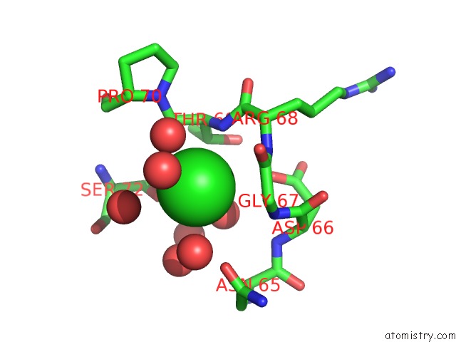
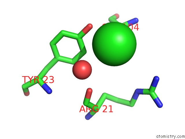
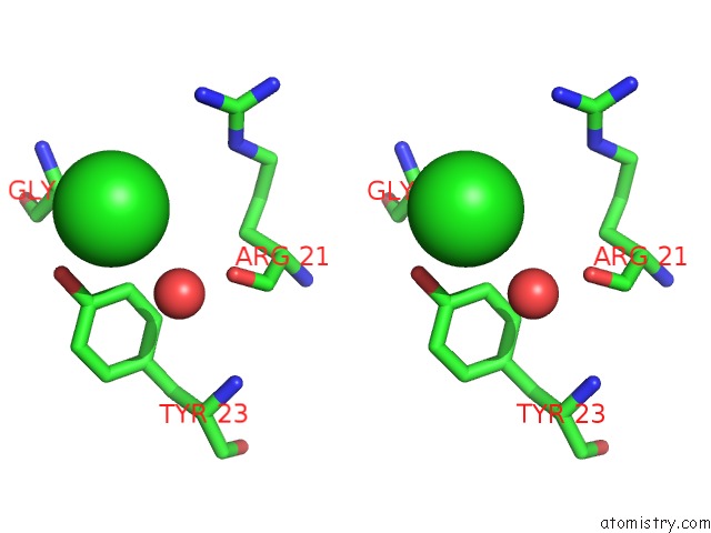
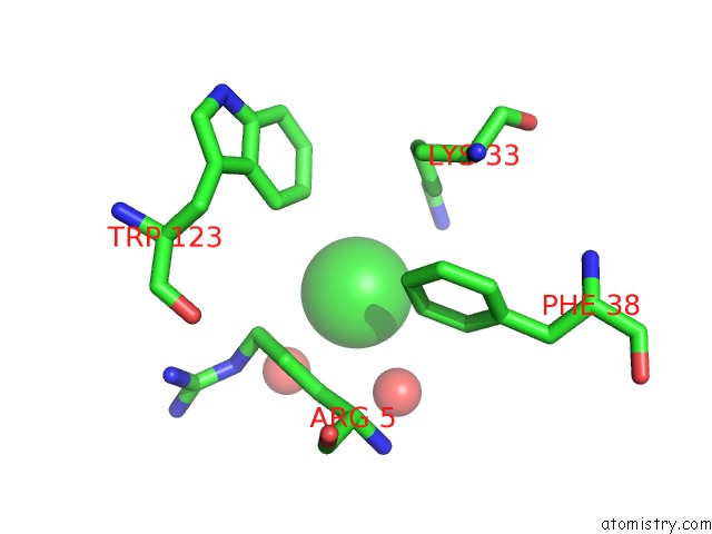
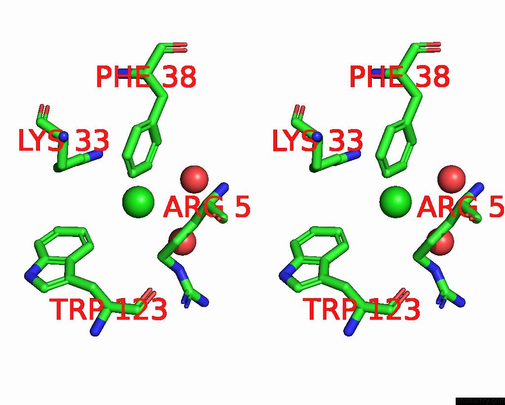
Chlorine binding site 1 out of 5 in 3ww6
Go back to
Chlorine binding site 1 out
of 5 in the Crystal Structure of Hen Egg White Lysozyme Mutant N46D/D52S
Mono view
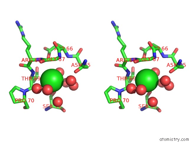
Stereo pair view
Mono view
Stereo pair view
A full contact list of Chlorine with other atoms in the Cl binding
site number 1 of Crystal Structure of Hen Egg White Lysozyme Mutant N46D/D52S within 5.0Å range:
|
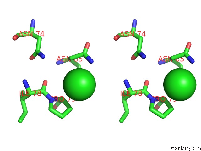
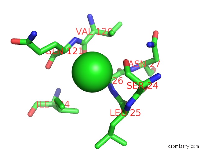
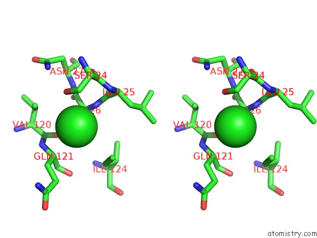
Chlorine binding site 2 out of 5 in 3ww6
Go back to
Chlorine binding site 2 out
of 5 in the Crystal Structure of Hen Egg White Lysozyme Mutant N46D/D52S
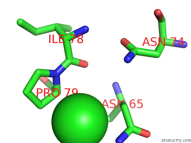
Mono view
Stereo pair view
Mono view
Stereo pair view
A full contact list of Chlorine with other atoms in the Cl binding
site number 2 of Crystal Structure of Hen Egg White Lysozyme Mutant N46D/D52S within 5.0Å range:
|
Chlorine binding site 3 out of 5 in 3ww6
Go back to
Chlorine binding site 3 out
of 5 in the Crystal Structure of Hen Egg White Lysozyme Mutant N46D/D52S
Mono view
Stereo pair view
Mono view
Stereo pair view
A full contact list of Chlorine with other atoms in the Cl binding
site number 3 of Crystal Structure of Hen Egg White Lysozyme Mutant N46D/D52S within 5.0Å range:
|
Chlorine binding site 4 out of 5 in 3ww6
Go back to
Chlorine binding site 4 out
of 5 in the Crystal Structure of Hen Egg White Lysozyme Mutant N46D/D52S
Mono view
Stereo pair view
Mono view
Stereo pair view
A full contact list of Chlorine with other atoms in the Cl binding
site number 4 of Crystal Structure of Hen Egg White Lysozyme Mutant N46D/D52S within 5.0Å range:
|
Chlorine binding site 5 out of 5 in 3ww6
Go back to
Chlorine binding site 5 out
of 5 in the Crystal Structure of Hen Egg White Lysozyme Mutant N46D/D52S
Mono view
Stereo pair view
Mono view
Stereo pair view
A full contact list of Chlorine with other atoms in the Cl binding
site number 5 of Crystal Structure of Hen Egg White Lysozyme Mutant N46D/D52S within 5.0Å range:
|
Reference:
Y.Abe,
M.Kubota,
S.Takazaki,
Y.Ito,
H.Yamamoto,
D.Kang,
T.Ueda,
T.Imoto.
Effect on Catalysis By Replacement of Catalytic Residue From Hen Egg White Lysozyme to Venerupis Philippinarum Lysozyme Protein Sci. 2016.
ISSN: ESSN 1469-896X
PubMed: 27291073
DOI: 10.1002/PRO.2966
Page generated: Sun Jul 21 07:49:19 2024
ISSN: ESSN 1469-896X
PubMed: 27291073
DOI: 10.1002/PRO.2966
Last articles
Zn in 9J0NZn in 9J0O
Zn in 9J0P
Zn in 9FJX
Zn in 9EKB
Zn in 9C0F
Zn in 9CAH
Zn in 9CH0
Zn in 9CH3
Zn in 9CH1