Chlorine »
PDB 3zh8-3zoj »
3zmh »
Chlorine in PDB 3zmh: Structure of E.Coli Rhomboid Protease Glpg in Complex with Monobactam L62
Enzymatic activity of Structure of E.Coli Rhomboid Protease Glpg in Complex with Monobactam L62
All present enzymatic activity of Structure of E.Coli Rhomboid Protease Glpg in Complex with Monobactam L62:
3.4.21.105;
3.4.21.105;
Protein crystallography data
The structure of Structure of E.Coli Rhomboid Protease Glpg in Complex with Monobactam L62, PDB code: 3zmh
was solved by
K.R.Vinothkumar,
O.A.Pierrat,
J.M.Large,
M.Freeman,
with X-Ray Crystallography technique. A brief refinement statistics is given in the table below:
| Resolution Low / High (Å) | 76.62 / 2.30 |
| Space group | H 3 2 |
| Cell size a, b, c (Å), α, β, γ (°) | 110.150, 110.150, 128.630, 90.00, 90.00, 120.00 |
| R / Rfree (%) | 18.8 / 22.9 |
Chlorine Binding Sites:
The binding sites of Chlorine atom in the Structure of E.Coli Rhomboid Protease Glpg in Complex with Monobactam L62
(pdb code 3zmh). This binding sites where shown within
5.0 Angstroms radius around Chlorine atom.
In total 2 binding sites of Chlorine where determined in the Structure of E.Coli Rhomboid Protease Glpg in Complex with Monobactam L62, PDB code: 3zmh:
Jump to Chlorine binding site number: 1; 2;
In total 2 binding sites of Chlorine where determined in the Structure of E.Coli Rhomboid Protease Glpg in Complex with Monobactam L62, PDB code: 3zmh:
Jump to Chlorine binding site number: 1; 2;
Chlorine binding site 1 out of 2 in 3zmh
Go back to
Chlorine binding site 1 out
of 2 in the Structure of E.Coli Rhomboid Protease Glpg in Complex with Monobactam L62

Mono view
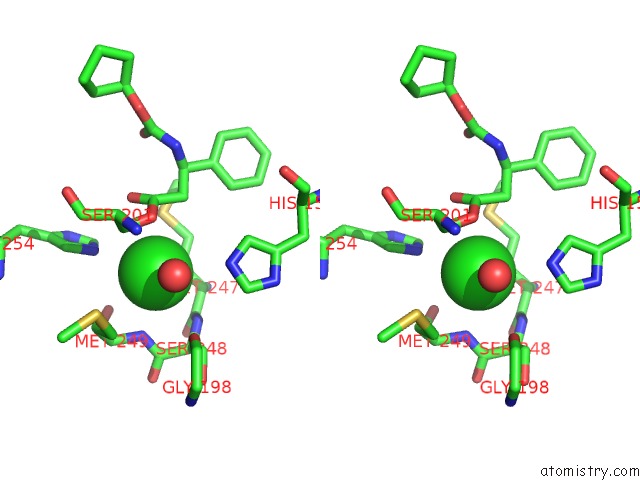
Stereo pair view
Mono view
Stereo pair view
A full contact list of Chlorine with other atoms in the Cl binding
site number 1 of Structure of E.Coli Rhomboid Protease Glpg in Complex with Monobactam L62 within 5.0Å range:
|
Chlorine binding site 2 out of 2 in 3zmh
Go back to
Chlorine binding site 2 out
of 2 in the Structure of E.Coli Rhomboid Protease Glpg in Complex with Monobactam L62

Mono view
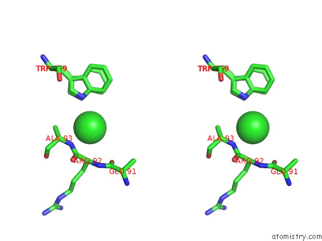
Stereo pair view
Mono view
Stereo pair view
A full contact list of Chlorine with other atoms in the Cl binding
site number 2 of Structure of E.Coli Rhomboid Protease Glpg in Complex with Monobactam L62 within 5.0Å range:
|
Reference:
K.R.Vinothkumar,
O.A.Pierrat,
J.M.Large,
M.Freeman.
Structure of Rhomboid Protease in Complex with Beta-Lactam Inhibitors Defines the S2' Cavity. Structure V. 21 1051 2013.
ISSN: ISSN 1878-4186
PubMed: 23665170
DOI: 10.1016/J.STR.2013.03.013
Page generated: Sun Jul 21 08:10:50 2024
ISSN: ISSN 1878-4186
PubMed: 23665170
DOI: 10.1016/J.STR.2013.03.013
Last articles
Zn in 9JYWZn in 9IR4
Zn in 9IR3
Zn in 9GMX
Zn in 9GMW
Zn in 9JEJ
Zn in 9ERF
Zn in 9ERE
Zn in 9EGV
Zn in 9EGW