Chlorine »
PDB 3zot-3zx2 »
3zsh »
Chlorine in PDB 3zsh: X-Ray Structure of P38ALPHA Bound to Scio-469
Enzymatic activity of X-Ray Structure of P38ALPHA Bound to Scio-469
All present enzymatic activity of X-Ray Structure of P38ALPHA Bound to Scio-469:
2.7.11.24;
2.7.11.24;
Protein crystallography data
The structure of X-Ray Structure of P38ALPHA Bound to Scio-469, PDB code: 3zsh
was solved by
R.Azevedo,
M.Van Zeeland,
H.Raaijmakers,
B.Kazemier,
A.Oubrie,
with X-Ray Crystallography technique. A brief refinement statistics is given in the table below:
| Resolution Low / High (Å) | 37.15 / 2.05 |
| Space group | P 21 21 21 |
| Cell size a, b, c (Å), α, β, γ (°) | 68.720, 70.080, 74.290, 90.00, 90.00, 90.00 |
| R / Rfree (%) | 20.5 / 25.5 |
Other elements in 3zsh:
The structure of X-Ray Structure of P38ALPHA Bound to Scio-469 also contains other interesting chemical elements:
| Fluorine | (F) | 1 atom |
Chlorine Binding Sites:
The binding sites of Chlorine atom in the X-Ray Structure of P38ALPHA Bound to Scio-469
(pdb code 3zsh). This binding sites where shown within
5.0 Angstroms radius around Chlorine atom.
In total only one binding site of Chlorine was determined in the X-Ray Structure of P38ALPHA Bound to Scio-469, PDB code: 3zsh:
In total only one binding site of Chlorine was determined in the X-Ray Structure of P38ALPHA Bound to Scio-469, PDB code: 3zsh:
Chlorine binding site 1 out of 1 in 3zsh
Go back to
Chlorine binding site 1 out
of 1 in the X-Ray Structure of P38ALPHA Bound to Scio-469
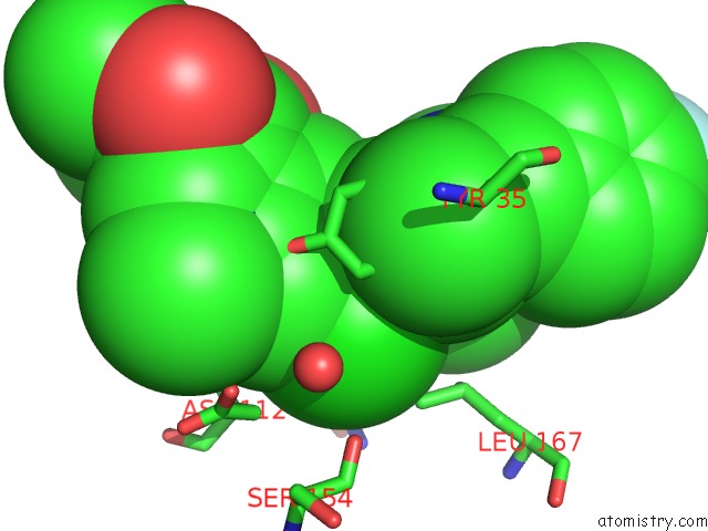
Mono view
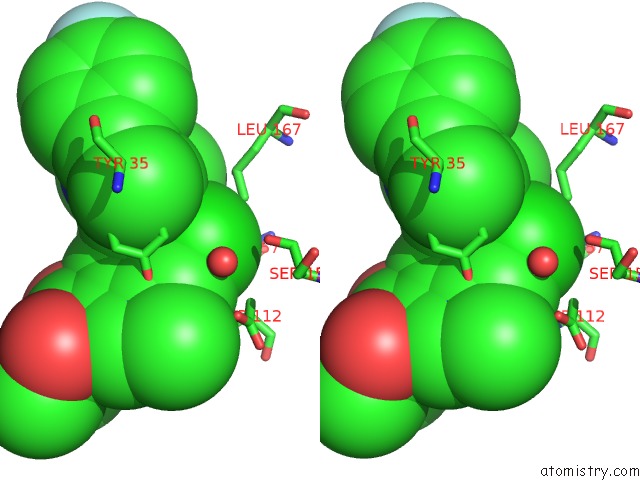
Stereo pair view
Mono view
Stereo pair view
A full contact list of Chlorine with other atoms in the Cl binding
site number 1 of X-Ray Structure of P38ALPHA Bound to Scio-469 within 5.0Å range:
|
Reference:
R.Azevedo,
M.Van Zeeland,
H.Raaijmakers,
B.Kazemier,
J.De Vlieg,
A.Oubrie.
X-Ray Structure of P38 Alpha Bound to Tak-715: Comparison with Three Classic Inhibitors. Acta Crystallogr. D Biol. V. 68 1041 2012CRYSTALLOGR..
ISSN: ESSN 1399-0047
PubMed: 22868770
DOI: 10.1107/S090744491201997X
Page generated: Sun Jul 21 08:28:42 2024
ISSN: ESSN 1399-0047
PubMed: 22868770
DOI: 10.1107/S090744491201997X
Last articles
Zn in 9J0NZn in 9J0O
Zn in 9J0P
Zn in 9FJX
Zn in 9EKB
Zn in 9C0F
Zn in 9CAH
Zn in 9CH0
Zn in 9CH3
Zn in 9CH1