Chlorine »
PDB 3zx3-4a7i »
4a6w »
Chlorine in PDB 4a6w: X-Ray Structures of Oxazole Hydroxamate Ecmetap-Mn Complexes
Enzymatic activity of X-Ray Structures of Oxazole Hydroxamate Ecmetap-Mn Complexes
All present enzymatic activity of X-Ray Structures of Oxazole Hydroxamate Ecmetap-Mn Complexes:
3.4.11.18;
3.4.11.18;
Protein crystallography data
The structure of X-Ray Structures of Oxazole Hydroxamate Ecmetap-Mn Complexes, PDB code: 4a6w
was solved by
F.Huguet,
A.Melet,
R.Alvesdesousa,
A.Lieutaud,
J.Chevalier,
P.Deschamps,
A.Tomas,
N.Leulliot,
J.M.Pages,
I.Artaud,
with X-Ray Crystallography technique. A brief refinement statistics is given in the table below:
| Resolution Low / High (Å) | 31.25 / 1.46 |
| Space group | P 1 21 1 |
| Cell size a, b, c (Å), α, β, γ (°) | 39.350, 62.910, 52.600, 90.00, 109.40, 90.00 |
| R / Rfree (%) | 15.296 / 22.623 |
Other elements in 4a6w:
The structure of X-Ray Structures of Oxazole Hydroxamate Ecmetap-Mn Complexes also contains other interesting chemical elements:
| Manganese | (Mn) | 2 atoms |
Chlorine Binding Sites:
The binding sites of Chlorine atom in the X-Ray Structures of Oxazole Hydroxamate Ecmetap-Mn Complexes
(pdb code 4a6w). This binding sites where shown within
5.0 Angstroms radius around Chlorine atom.
In total only one binding site of Chlorine was determined in the X-Ray Structures of Oxazole Hydroxamate Ecmetap-Mn Complexes, PDB code: 4a6w:
In total only one binding site of Chlorine was determined in the X-Ray Structures of Oxazole Hydroxamate Ecmetap-Mn Complexes, PDB code: 4a6w:
Chlorine binding site 1 out of 1 in 4a6w
Go back to
Chlorine binding site 1 out
of 1 in the X-Ray Structures of Oxazole Hydroxamate Ecmetap-Mn Complexes
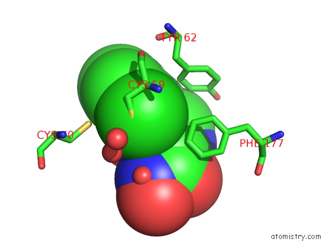
Mono view
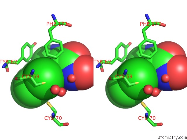
Stereo pair view
Mono view
Stereo pair view
A full contact list of Chlorine with other atoms in the Cl binding
site number 1 of X-Ray Structures of Oxazole Hydroxamate Ecmetap-Mn Complexes within 5.0Å range:
|
Reference:
F.Huguet,
A.Melet,
R.Alves De Sousa,
A.Lieutaud,
J.Chevalier,
L.Maigre,
P.Deschamps,
A.Tomas,
N.Leulliot,
J.M.Pages,
I.Artaud.
Hydroxamic Acids As Potent Inhibitors of Fe(II) and Mn(II) E. Coli Methionine Aminopeptidase: Biological Activities and X-Ray Structures of Oxazole Hydroxamate-Ecmetap-Mn Complexes. Chemmedchem V. 7 1020 2012.
ISSN: ISSN 1860-7179
PubMed: 22489069
DOI: 10.1002/CMDC.201200076
Page generated: Sun Jul 21 08:54:40 2024
ISSN: ISSN 1860-7179
PubMed: 22489069
DOI: 10.1002/CMDC.201200076
Last articles
Ca in 5NMRCa in 5NN9
Ca in 5NM8
Ca in 5NH8
Ca in 5NL7
Ca in 5NIN
Ca in 5NGQ
Ca in 5NH5
Ca in 5NGY
Ca in 5NG1