Chlorine »
PDB 4aj2-4aqd »
4ap6 »
Chlorine in PDB 4ap6: Crystal Structure of Human POFUT2 E54A Mutant in Complex with Gdp- Fucose
Enzymatic activity of Crystal Structure of Human POFUT2 E54A Mutant in Complex with Gdp- Fucose
All present enzymatic activity of Crystal Structure of Human POFUT2 E54A Mutant in Complex with Gdp- Fucose:
2.4.1.221;
2.4.1.221;
Protein crystallography data
The structure of Crystal Structure of Human POFUT2 E54A Mutant in Complex with Gdp- Fucose, PDB code: 4ap6
was solved by
C.Chen,
J.J.Keusch,
D.Klein,
D.Hess,
J.Hofsteenge,
H.Gut,
with X-Ray Crystallography technique. A brief refinement statistics is given in the table below:
| Resolution Low / High (Å) | 39.686 / 3.401 |
| Space group | P 32 2 1 |
| Cell size a, b, c (Å), α, β, γ (°) | 153.010, 153.010, 185.680, 90.00, 90.00, 120.00 |
| R / Rfree (%) | 19.24 / 23.8 |
Chlorine Binding Sites:
The binding sites of Chlorine atom in the Crystal Structure of Human POFUT2 E54A Mutant in Complex with Gdp- Fucose
(pdb code 4ap6). This binding sites where shown within
5.0 Angstroms radius around Chlorine atom.
In total 4 binding sites of Chlorine where determined in the Crystal Structure of Human POFUT2 E54A Mutant in Complex with Gdp- Fucose, PDB code: 4ap6:
Jump to Chlorine binding site number: 1; 2; 3; 4;
In total 4 binding sites of Chlorine where determined in the Crystal Structure of Human POFUT2 E54A Mutant in Complex with Gdp- Fucose, PDB code: 4ap6:
Jump to Chlorine binding site number: 1; 2; 3; 4;
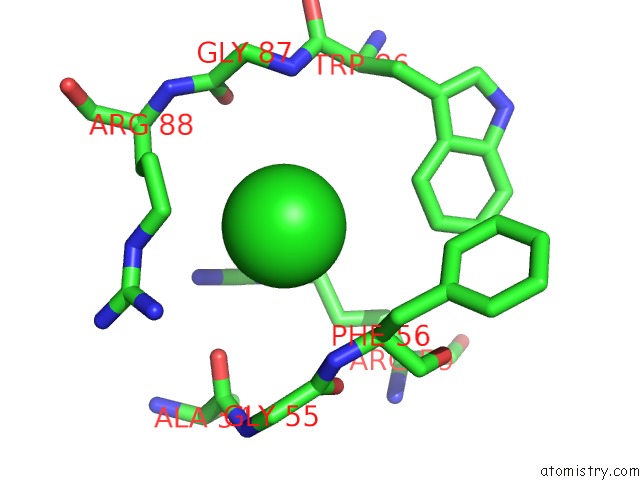
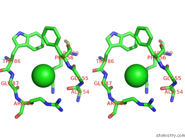
Chlorine binding site 1 out of 4 in 4ap6
Go back to
Chlorine binding site 1 out
of 4 in the Crystal Structure of Human POFUT2 E54A Mutant in Complex with Gdp- Fucose
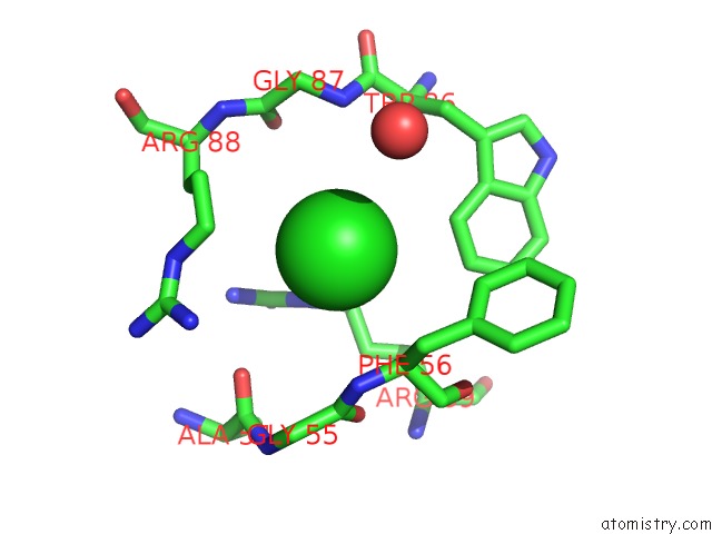
Mono view
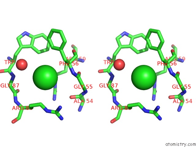
Stereo pair view
Mono view
Stereo pair view
A full contact list of Chlorine with other atoms in the Cl binding
site number 1 of Crystal Structure of Human POFUT2 E54A Mutant in Complex with Gdp- Fucose within 5.0Å range:
|
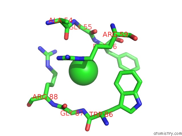
Chlorine binding site 2 out of 4 in 4ap6
Go back to
Chlorine binding site 2 out
of 4 in the Crystal Structure of Human POFUT2 E54A Mutant in Complex with Gdp- Fucose
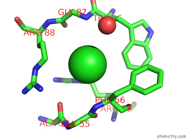
Mono view
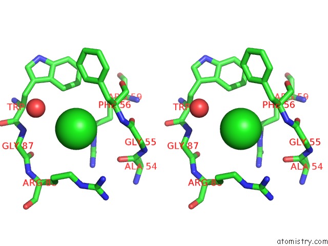
Stereo pair view
Mono view
Stereo pair view
A full contact list of Chlorine with other atoms in the Cl binding
site number 2 of Crystal Structure of Human POFUT2 E54A Mutant in Complex with Gdp- Fucose within 5.0Å range:
|
Chlorine binding site 3 out of 4 in 4ap6
Go back to
Chlorine binding site 3 out
of 4 in the Crystal Structure of Human POFUT2 E54A Mutant in Complex with Gdp- Fucose
Mono view
Stereo pair view
Mono view
Stereo pair view
A full contact list of Chlorine with other atoms in the Cl binding
site number 3 of Crystal Structure of Human POFUT2 E54A Mutant in Complex with Gdp- Fucose within 5.0Å range:
|
Chlorine binding site 4 out of 4 in 4ap6
Go back to
Chlorine binding site 4 out
of 4 in the Crystal Structure of Human POFUT2 E54A Mutant in Complex with Gdp- Fucose
Mono view

Stereo pair view
Mono view
Stereo pair view
A full contact list of Chlorine with other atoms in the Cl binding
site number 4 of Crystal Structure of Human POFUT2 E54A Mutant in Complex with Gdp- Fucose within 5.0Å range:
|
Reference:
C.I.Chen,
J.J.Keusch,
D.Klein,
D.Hess,
J.Hofsteenge,
H.Gut.
Structure of Human POFUT2: Insights Into Thrombospondin Type 1 Repeat Fold and O-Fucosylation. Embo J. V. 31 3183 2012.
ISSN: ISSN 0261-4189
PubMed: 22588082
DOI: 10.1038/EMBOJ.2012.143
Page generated: Sun Jul 21 09:18:49 2024
ISSN: ISSN 0261-4189
PubMed: 22588082
DOI: 10.1038/EMBOJ.2012.143
Last articles
Zn in 9J0NZn in 9J0O
Zn in 9J0P
Zn in 9FJX
Zn in 9EKB
Zn in 9C0F
Zn in 9CAH
Zn in 9CH0
Zn in 9CH3
Zn in 9CH1