Chlorine »
PDB 4ced-4ck4 »
4cf6 »
Chlorine in PDB 4cf6: Crystal Structure of the Complex of the P187S Variant of Human Nad(P)H:Quinone Oxidoreductase with Cibacron Blue at 2.7 A Resolution
Enzymatic activity of Crystal Structure of the Complex of the P187S Variant of Human Nad(P)H:Quinone Oxidoreductase with Cibacron Blue at 2.7 A Resolution
All present enzymatic activity of Crystal Structure of the Complex of the P187S Variant of Human Nad(P)H:Quinone Oxidoreductase with Cibacron Blue at 2.7 A Resolution:
1.6.5.2;
1.6.5.2;
Protein crystallography data
The structure of Crystal Structure of the Complex of the P187S Variant of Human Nad(P)H:Quinone Oxidoreductase with Cibacron Blue at 2.7 A Resolution, PDB code: 4cf6
was solved by
W.D.Lienhart,
V.Gudipati,
M.K.Uhl,
A.Binter,
S.Pulido,
R.Saf,
K.Zangger,
K.Gruber,
P.Macheroux,
with X-Ray Crystallography technique. A brief refinement statistics is given in the table below:
| Resolution Low / High (Å) | 46.217 / 2.69 |
| Space group | I 2 2 2 |
| Cell size a, b, c (Å), α, β, γ (°) | 104.167, 104.563, 118.566, 90.00, 90.00, 90.00 |
| R / Rfree (%) | 18.47 / 20.74 |
Chlorine Binding Sites:
The binding sites of Chlorine atom in the Crystal Structure of the Complex of the P187S Variant of Human Nad(P)H:Quinone Oxidoreductase with Cibacron Blue at 2.7 A Resolution
(pdb code 4cf6). This binding sites where shown within
5.0 Angstroms radius around Chlorine atom.
In total 2 binding sites of Chlorine where determined in the Crystal Structure of the Complex of the P187S Variant of Human Nad(P)H:Quinone Oxidoreductase with Cibacron Blue at 2.7 A Resolution, PDB code: 4cf6:
Jump to Chlorine binding site number: 1; 2;
In total 2 binding sites of Chlorine where determined in the Crystal Structure of the Complex of the P187S Variant of Human Nad(P)H:Quinone Oxidoreductase with Cibacron Blue at 2.7 A Resolution, PDB code: 4cf6:
Jump to Chlorine binding site number: 1; 2;
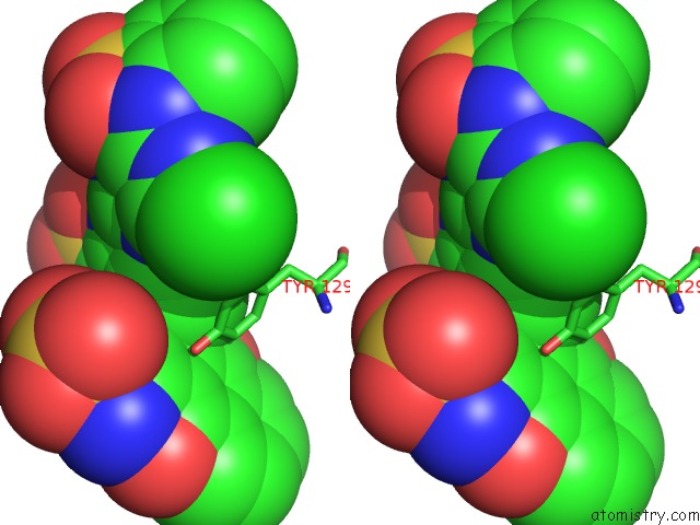
Chlorine binding site 1 out of 2 in 4cf6
Go back to
Chlorine binding site 1 out
of 2 in the Crystal Structure of the Complex of the P187S Variant of Human Nad(P)H:Quinone Oxidoreductase with Cibacron Blue at 2.7 A Resolution
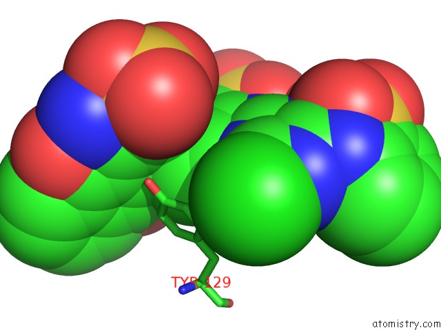
Mono view
Stereo pair view
Mono view
Stereo pair view
A full contact list of Chlorine with other atoms in the Cl binding
site number 1 of Crystal Structure of the Complex of the P187S Variant of Human Nad(P)H:Quinone Oxidoreductase with Cibacron Blue at 2.7 A Resolution within 5.0Å range:
|
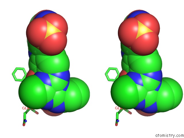
Chlorine binding site 2 out of 2 in 4cf6
Go back to
Chlorine binding site 2 out
of 2 in the Crystal Structure of the Complex of the P187S Variant of Human Nad(P)H:Quinone Oxidoreductase with Cibacron Blue at 2.7 A Resolution
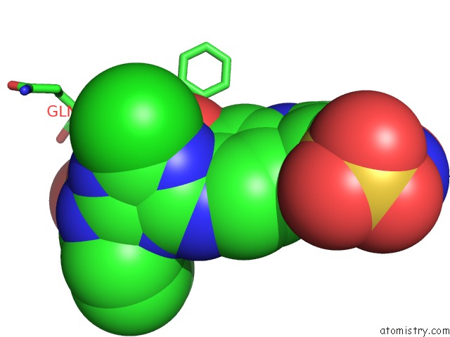
Mono view
Stereo pair view
Mono view
Stereo pair view
A full contact list of Chlorine with other atoms in the Cl binding
site number 2 of Crystal Structure of the Complex of the P187S Variant of Human Nad(P)H:Quinone Oxidoreductase with Cibacron Blue at 2.7 A Resolution within 5.0Å range:
|
Reference:
W.D.Lienhart,
V.Gudipati,
M.K.Uhl,
A.Binter,
S.Pulido,
R.Saf,
K.Zangger,
K.Gruber,
P.Macheroux.
Collapse of the Native Structure By A Single Amino Acid Exchange in Human Nad(P)H:Quinone Oxidoreductase (NQO1). Febs J. V. 281 4691 2014.
ISSN: ISSN 1742-464X
PubMed: 25143260
DOI: 10.1111/FEBS.12975
Page generated: Sun Jul 21 11:10:47 2024
ISSN: ISSN 1742-464X
PubMed: 25143260
DOI: 10.1111/FEBS.12975
Last articles
Zn in 9J0NZn in 9J0O
Zn in 9J0P
Zn in 9FJX
Zn in 9EKB
Zn in 9C0F
Zn in 9CAH
Zn in 9CH0
Zn in 9CH3
Zn in 9CH1