Chlorine »
PDB 4ced-4ck4 »
4ciy »
Chlorine in PDB 4ciy: Crystal Structure of Mycobacterium Tuberculosis Type 2 Dehydroquinase in Complex with (1R,4R,5R)-1,4,5-Trihydroxy-3-((1R)-1-Hydroxy-2- Phenyl)Ethylcyclohex-2-En-1-Carboxylic Acid
Enzymatic activity of Crystal Structure of Mycobacterium Tuberculosis Type 2 Dehydroquinase in Complex with (1R,4R,5R)-1,4,5-Trihydroxy-3-((1R)-1-Hydroxy-2- Phenyl)Ethylcyclohex-2-En-1-Carboxylic Acid
All present enzymatic activity of Crystal Structure of Mycobacterium Tuberculosis Type 2 Dehydroquinase in Complex with (1R,4R,5R)-1,4,5-Trihydroxy-3-((1R)-1-Hydroxy-2- Phenyl)Ethylcyclohex-2-En-1-Carboxylic Acid:
4.2.1.10;
4.2.1.10;
Protein crystallography data
The structure of Crystal Structure of Mycobacterium Tuberculosis Type 2 Dehydroquinase in Complex with (1R,4R,5R)-1,4,5-Trihydroxy-3-((1R)-1-Hydroxy-2- Phenyl)Ethylcyclohex-2-En-1-Carboxylic Acid, PDB code: 4ciy
was solved by
J.M.Otero,
A.L.Llamas-Saiz,
H.Lamb,
A.R.Hawkins,
B.Blanco,
A.Sedes,
A.Peon,
C.Gonzalez-Bello,
M.J.Van Raaij,
with X-Ray Crystallography technique. A brief refinement statistics is given in the table below:
| Resolution Low / High (Å) | 38.08 / 2.10 |
| Space group | F 2 3 |
| Cell size a, b, c (Å), α, β, γ (°) | 126.205, 126.205, 126.205, 90.00, 90.00, 90.00 |
| R / Rfree (%) | 14.549 / 19.754 |
Other elements in 4ciy:
The structure of Crystal Structure of Mycobacterium Tuberculosis Type 2 Dehydroquinase in Complex with (1R,4R,5R)-1,4,5-Trihydroxy-3-((1R)-1-Hydroxy-2- Phenyl)Ethylcyclohex-2-En-1-Carboxylic Acid also contains other interesting chemical elements:
| Sodium | (Na) | 3 atoms |
Chlorine Binding Sites:
The binding sites of Chlorine atom in the Crystal Structure of Mycobacterium Tuberculosis Type 2 Dehydroquinase in Complex with (1R,4R,5R)-1,4,5-Trihydroxy-3-((1R)-1-Hydroxy-2- Phenyl)Ethylcyclohex-2-En-1-Carboxylic Acid
(pdb code 4ciy). This binding sites where shown within
5.0 Angstroms radius around Chlorine atom.
In total only one binding site of Chlorine was determined in the Crystal Structure of Mycobacterium Tuberculosis Type 2 Dehydroquinase in Complex with (1R,4R,5R)-1,4,5-Trihydroxy-3-((1R)-1-Hydroxy-2- Phenyl)Ethylcyclohex-2-En-1-Carboxylic Acid, PDB code: 4ciy:
In total only one binding site of Chlorine was determined in the Crystal Structure of Mycobacterium Tuberculosis Type 2 Dehydroquinase in Complex with (1R,4R,5R)-1,4,5-Trihydroxy-3-((1R)-1-Hydroxy-2- Phenyl)Ethylcyclohex-2-En-1-Carboxylic Acid, PDB code: 4ciy:
Chlorine binding site 1 out of 1 in 4ciy
Go back to
Chlorine binding site 1 out
of 1 in the Crystal Structure of Mycobacterium Tuberculosis Type 2 Dehydroquinase in Complex with (1R,4R,5R)-1,4,5-Trihydroxy-3-((1R)-1-Hydroxy-2- Phenyl)Ethylcyclohex-2-En-1-Carboxylic Acid
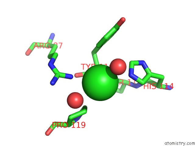
Mono view
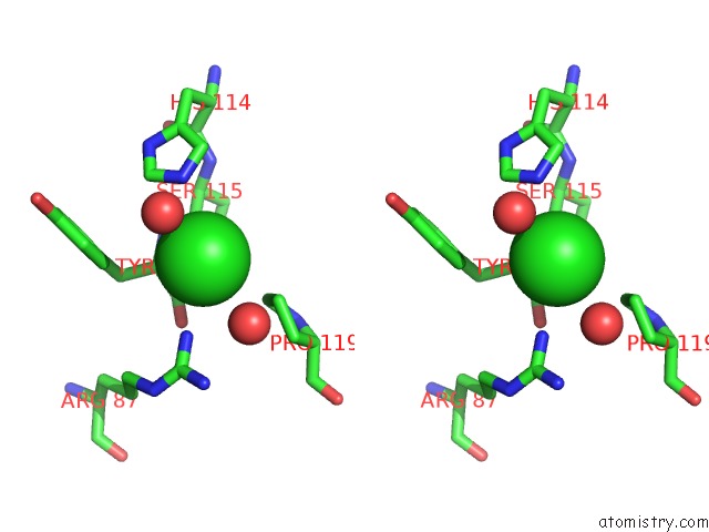
Stereo pair view
Mono view
Stereo pair view
A full contact list of Chlorine with other atoms in the Cl binding
site number 1 of Crystal Structure of Mycobacterium Tuberculosis Type 2 Dehydroquinase in Complex with (1R,4R,5R)-1,4,5-Trihydroxy-3-((1R)-1-Hydroxy-2- Phenyl)Ethylcyclohex-2-En-1-Carboxylic Acid within 5.0Å range:
|
Reference:
B.Blanco,
A.Sedes,
A.Peon,
J.M.Otero,
M.J.Van Raaij,
P.Thompson,
A.R.Hawkins,
C.Gonzalez-Bello.
Exploring the Water-Binding Pocket of the Type II Dehydroquinase Enzyme in the Structure-Based Design of Inhibitors. J.Med.Chem. V. 57 3494 2014.
ISSN: ISSN 0022-2623
PubMed: 24689821
DOI: 10.1021/JM500175Z
Page generated: Sun Jul 21 11:15:32 2024
ISSN: ISSN 0022-2623
PubMed: 24689821
DOI: 10.1021/JM500175Z
Last articles
Zn in 9MJ5Zn in 9HNW
Zn in 9G0L
Zn in 9FNE
Zn in 9DZN
Zn in 9E0I
Zn in 9D32
Zn in 9DAK
Zn in 8ZXC
Zn in 8ZUF