Chlorine »
PDB 4da5-4dhn »
4db3 »
Chlorine in PDB 4db3: 1.95 Angstrom Resolution Crystal Structure of N-Acetyl-D-Glucosamine Kinase From Vibrio Vulnificus.
Enzymatic activity of 1.95 Angstrom Resolution Crystal Structure of N-Acetyl-D-Glucosamine Kinase From Vibrio Vulnificus.
All present enzymatic activity of 1.95 Angstrom Resolution Crystal Structure of N-Acetyl-D-Glucosamine Kinase From Vibrio Vulnificus.:
2.7.1.59;
2.7.1.59;
Protein crystallography data
The structure of 1.95 Angstrom Resolution Crystal Structure of N-Acetyl-D-Glucosamine Kinase From Vibrio Vulnificus., PDB code: 4db3
was solved by
G.Minasov,
Z.Wawrzak,
O.Onopriyenko,
T.Skarina,
L.Papazisi,
A.Savchenko,
W.F.Anderson,
Center For Structural Genomics Of Infectious Diseases(Csgid),
with X-Ray Crystallography technique. A brief refinement statistics is given in the table below:
| Resolution Low / High (Å) | 29.45 / 1.95 |
| Space group | P 64 |
| Cell size a, b, c (Å), α, β, γ (°) | 80.400, 80.400, 110.418, 90.00, 90.00, 120.00 |
| R / Rfree (%) | 16.8 / 19.8 |
Other elements in 4db3:
The structure of 1.95 Angstrom Resolution Crystal Structure of N-Acetyl-D-Glucosamine Kinase From Vibrio Vulnificus. also contains other interesting chemical elements:
| Zinc | (Zn) | 1 atom |
Chlorine Binding Sites:
The binding sites of Chlorine atom in the 1.95 Angstrom Resolution Crystal Structure of N-Acetyl-D-Glucosamine Kinase From Vibrio Vulnificus.
(pdb code 4db3). This binding sites where shown within
5.0 Angstroms radius around Chlorine atom.
In total 3 binding sites of Chlorine where determined in the 1.95 Angstrom Resolution Crystal Structure of N-Acetyl-D-Glucosamine Kinase From Vibrio Vulnificus., PDB code: 4db3:
Jump to Chlorine binding site number: 1; 2; 3;
In total 3 binding sites of Chlorine where determined in the 1.95 Angstrom Resolution Crystal Structure of N-Acetyl-D-Glucosamine Kinase From Vibrio Vulnificus., PDB code: 4db3:
Jump to Chlorine binding site number: 1; 2; 3;
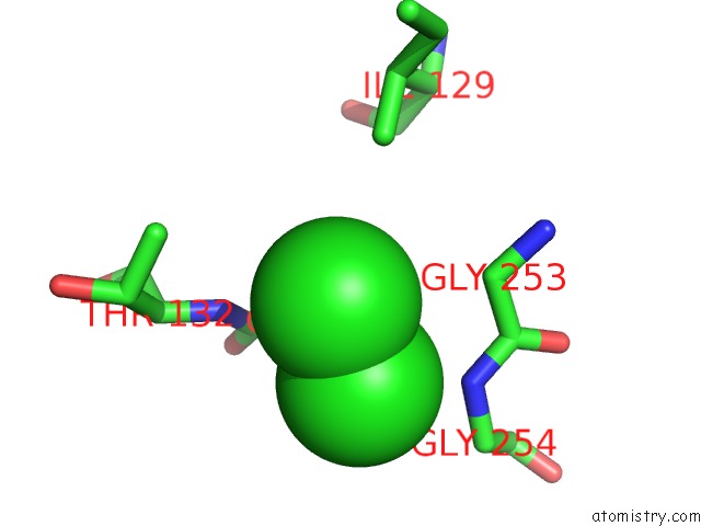
Chlorine binding site 1 out of 3 in 4db3
Go back to
Chlorine binding site 1 out
of 3 in the 1.95 Angstrom Resolution Crystal Structure of N-Acetyl-D-Glucosamine Kinase From Vibrio Vulnificus.
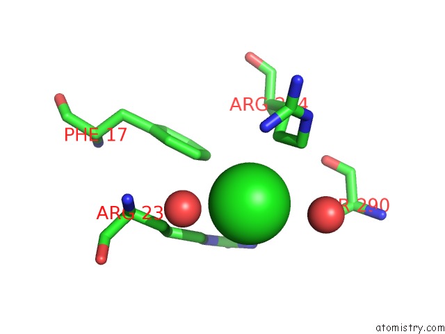
Mono view
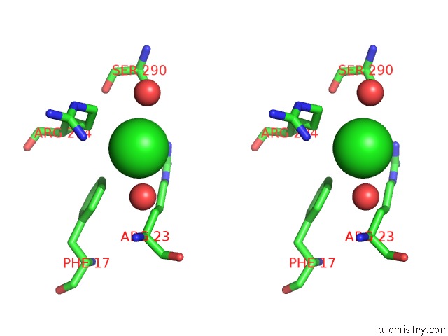
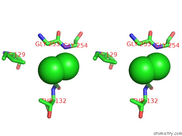
Stereo pair view
Mono view
Stereo pair view
A full contact list of Chlorine with other atoms in the Cl binding
site number 1 of 1.95 Angstrom Resolution Crystal Structure of N-Acetyl-D-Glucosamine Kinase From Vibrio Vulnificus. within 5.0Å range:
|
Chlorine binding site 2 out of 3 in 4db3
Go back to
Chlorine binding site 2 out
of 3 in the 1.95 Angstrom Resolution Crystal Structure of N-Acetyl-D-Glucosamine Kinase From Vibrio Vulnificus.
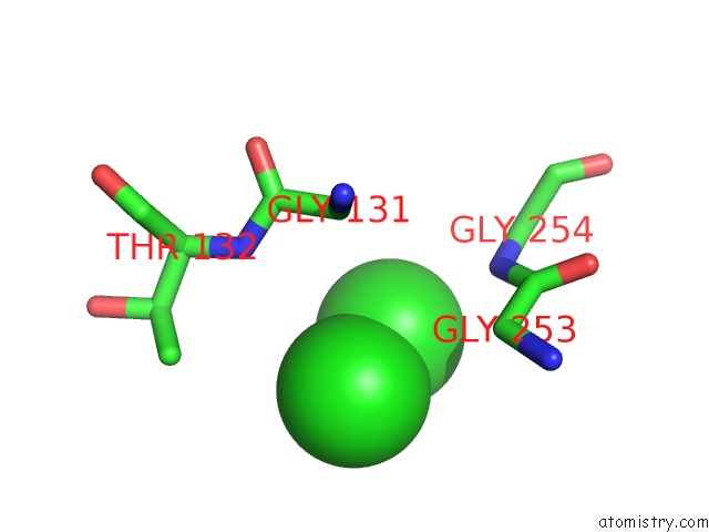
Mono view
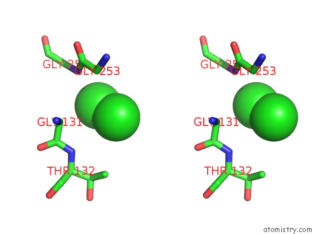
Stereo pair view
Mono view
Stereo pair view
A full contact list of Chlorine with other atoms in the Cl binding
site number 2 of 1.95 Angstrom Resolution Crystal Structure of N-Acetyl-D-Glucosamine Kinase From Vibrio Vulnificus. within 5.0Å range:
|
Chlorine binding site 3 out of 3 in 4db3
Go back to
Chlorine binding site 3 out
of 3 in the 1.95 Angstrom Resolution Crystal Structure of N-Acetyl-D-Glucosamine Kinase From Vibrio Vulnificus.
Mono view
Stereo pair view
Mono view
Stereo pair view
A full contact list of Chlorine with other atoms in the Cl binding
site number 3 of 1.95 Angstrom Resolution Crystal Structure of N-Acetyl-D-Glucosamine Kinase From Vibrio Vulnificus. within 5.0Å range:
|
Reference:
G.Minasov,
Z.Wawrzak,
O.Onopriyenko,
T.Skarina,
L.Papazisi,
A.Savchenko,
W.F.Anderson,
Center For Structural Genomics Of Infectious Diseases(Csgid).
1.95 Angstrom Resolution Crystal Structure of N-Acetyl-D-Glucosamine Kinase From Vibrio Vulnificus. To Be Published.
Page generated: Sun Jul 21 11:54:04 2024
Last articles
Ca in 5MNOCa in 5MNN
Ca in 5MKI
Ca in 5MNM
Ca in 5MNL
Ca in 5MNK
Ca in 5MNH
Ca in 5MNG
Ca in 5MNF
Ca in 5MNE