Chlorine »
PDB 4dho-4dq6 »
4dkl »
Chlorine in PDB 4dkl: Crystal Structure of the Mu-Opioid Receptor Bound to A Morphinan Antagonist
Enzymatic activity of Crystal Structure of the Mu-Opioid Receptor Bound to A Morphinan Antagonist
All present enzymatic activity of Crystal Structure of the Mu-Opioid Receptor Bound to A Morphinan Antagonist:
3.2.1.17;
3.2.1.17;
Protein crystallography data
The structure of Crystal Structure of the Mu-Opioid Receptor Bound to A Morphinan Antagonist, PDB code: 4dkl
was solved by
A.Manglik,
A.C.Kruse,
T.S.Kobilka,
F.S.Thian,
J.M.Mathiesen,
R.K.Sunahara,
L.Pardo,
W.I.Weis,
B.K.Kobilka,
S.Granier,
with X-Ray Crystallography technique. A brief refinement statistics is given in the table below:
| Resolution Low / High (Å) | 30.49 / 2.80 |
| Space group | C 1 2 1 |
| Cell size a, b, c (Å), α, β, γ (°) | 70.882, 174.730, 68.353, 90.00, 107.84, 90.00 |
| R / Rfree (%) | 23.3 / 27.5 |
Chlorine Binding Sites:
The binding sites of Chlorine atom in the Crystal Structure of the Mu-Opioid Receptor Bound to A Morphinan Antagonist
(pdb code 4dkl). This binding sites where shown within
5.0 Angstroms radius around Chlorine atom.
In total only one binding site of Chlorine was determined in the Crystal Structure of the Mu-Opioid Receptor Bound to A Morphinan Antagonist, PDB code: 4dkl:
In total only one binding site of Chlorine was determined in the Crystal Structure of the Mu-Opioid Receptor Bound to A Morphinan Antagonist, PDB code: 4dkl:
Chlorine binding site 1 out of 1 in 4dkl
Go back to
Chlorine binding site 1 out
of 1 in the Crystal Structure of the Mu-Opioid Receptor Bound to A Morphinan Antagonist
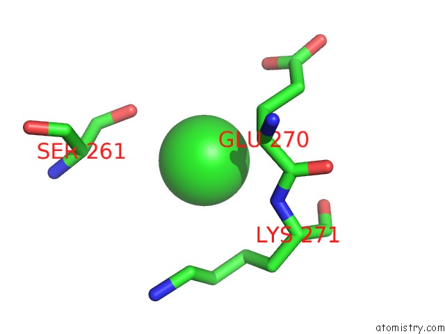
Mono view
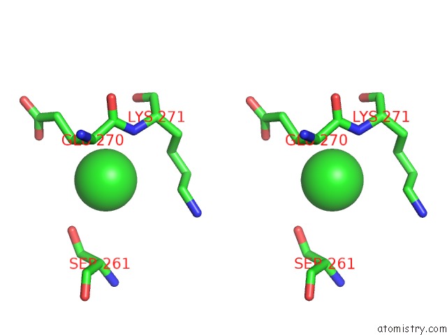
Stereo pair view
Mono view
Stereo pair view
A full contact list of Chlorine with other atoms in the Cl binding
site number 1 of Crystal Structure of the Mu-Opioid Receptor Bound to A Morphinan Antagonist within 5.0Å range:
|
Reference:
A.Manglik,
A.C.Kruse,
T.S.Kobilka,
F.S.Thian,
J.M.Mathiesen,
R.K.Sunahara,
L.Pardo,
W.I.Weis,
B.K.Kobilka,
S.Granier.
Crystal Structure of the {Mu}-Opioid Receptor Bound to A Morphinan Antagonist. Nature V. 485 321 2012.
ISSN: ISSN 0028-0836
PubMed: 22437502
DOI: 10.1038/NATURE10954
Page generated: Sun Jul 21 12:09:14 2024
ISSN: ISSN 0028-0836
PubMed: 22437502
DOI: 10.1038/NATURE10954
Last articles
Zn in 9J0NZn in 9J0O
Zn in 9J0P
Zn in 9FJX
Zn in 9EKB
Zn in 9C0F
Zn in 9CAH
Zn in 9CH0
Zn in 9CH3
Zn in 9CH1