Chlorine »
PDB 4e0j-4e9v »
4e7v »
Chlorine in PDB 4e7v: The Structure of R6 Bovine Insulin
Protein crystallography data
The structure of The Structure of R6 Bovine Insulin, PDB code: 4e7v
was solved by
P.Harris,
C.G.Frankaer,
M.V.Knudsen,
with X-Ray Crystallography technique. A brief refinement statistics is given in the table below:
| Resolution Low / High (Å) | 28.88 / 1.80 |
| Space group | H 3 1 |
| Cell size a, b, c (Å), α, β, γ (°) | 156.240, 156.240, 78.880, 90.00, 90.00, 120.00 |
| R / Rfree (%) | 20.9 / 27.2 |
Other elements in 4e7v:
The structure of The Structure of R6 Bovine Insulin also contains other interesting chemical elements:
| Zinc | (Zn) | 8 atoms |
Chlorine Binding Sites:
The binding sites of Chlorine atom in the The Structure of R6 Bovine Insulin
(pdb code 4e7v). This binding sites where shown within
5.0 Angstroms radius around Chlorine atom.
In total 8 binding sites of Chlorine where determined in the The Structure of R6 Bovine Insulin, PDB code: 4e7v:
Jump to Chlorine binding site number: 1; 2; 3; 4; 5; 6; 7; 8;
In total 8 binding sites of Chlorine where determined in the The Structure of R6 Bovine Insulin, PDB code: 4e7v:
Jump to Chlorine binding site number: 1; 2; 3; 4; 5; 6; 7; 8;
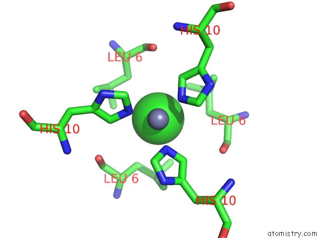
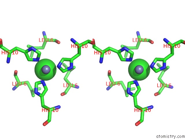
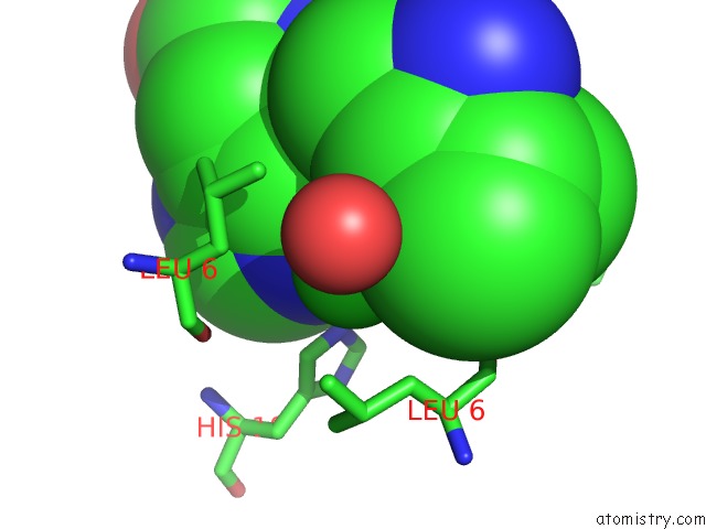
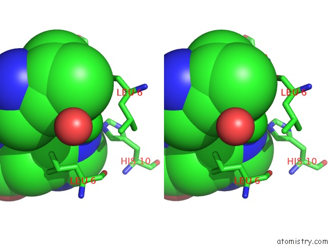
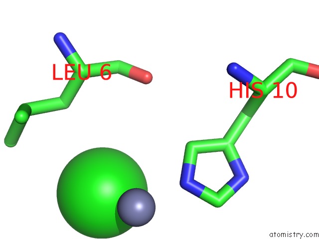
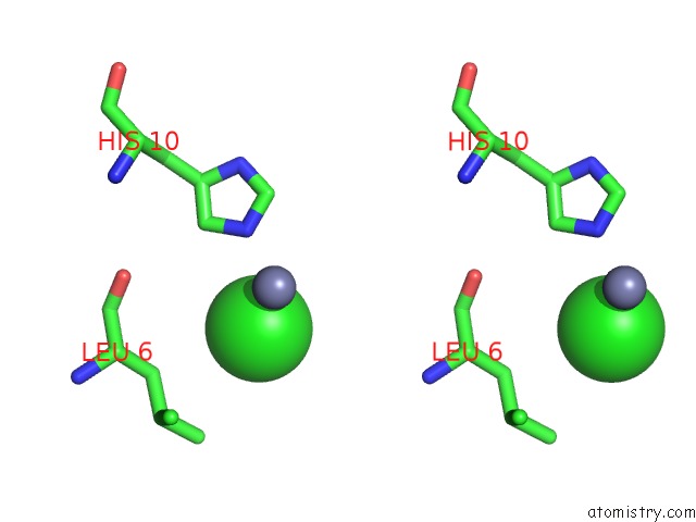
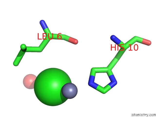
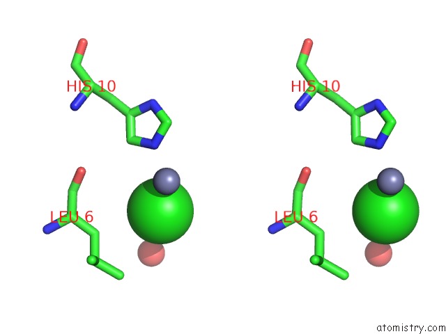
Chlorine binding site 1 out of 8 in 4e7v
Go back to
Chlorine binding site 1 out
of 8 in the The Structure of R6 Bovine Insulin
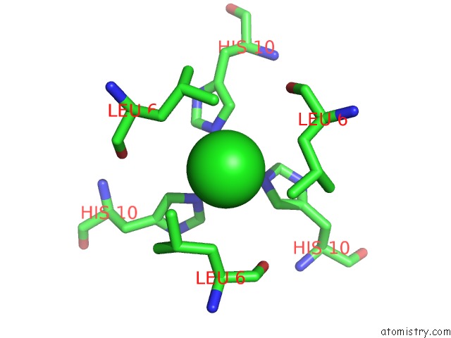
Mono view
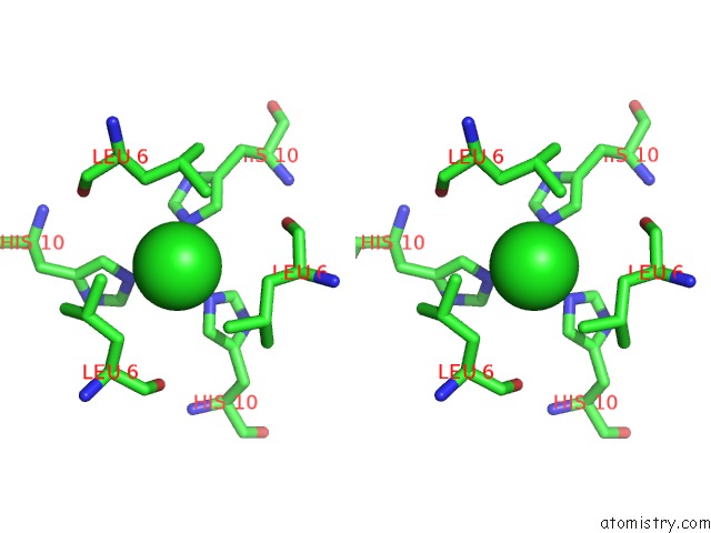
Stereo pair view
Mono view
Stereo pair view
A full contact list of Chlorine with other atoms in the Cl binding
site number 1 of The Structure of R6 Bovine Insulin within 5.0Å range:
|
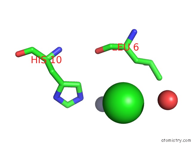
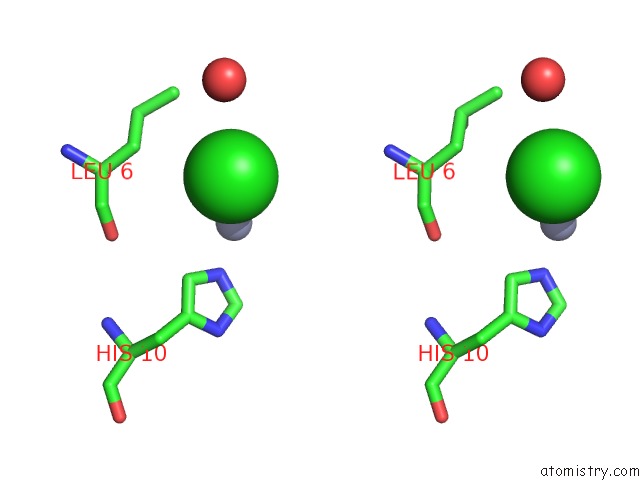
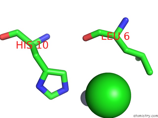
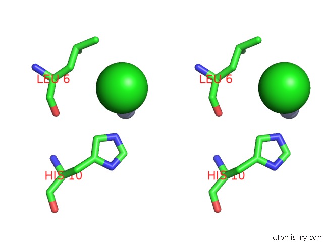
Chlorine binding site 2 out of 8 in 4e7v
Go back to
Chlorine binding site 2 out
of 8 in the The Structure of R6 Bovine Insulin
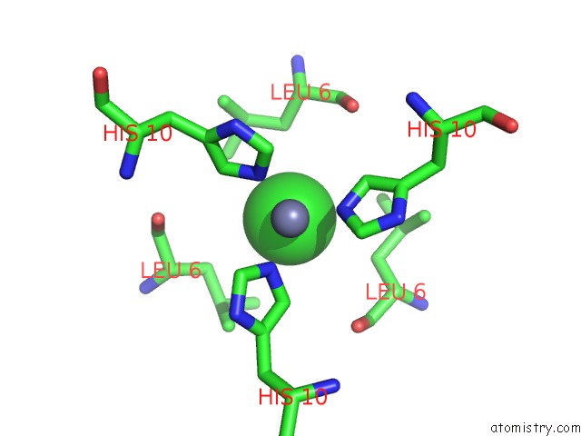
Mono view
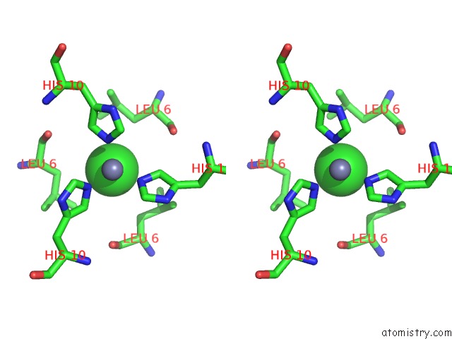
Stereo pair view
Mono view
Stereo pair view
A full contact list of Chlorine with other atoms in the Cl binding
site number 2 of The Structure of R6 Bovine Insulin within 5.0Å range:
|
Chlorine binding site 3 out of 8 in 4e7v
Go back to
Chlorine binding site 3 out
of 8 in the The Structure of R6 Bovine Insulin
Mono view
Stereo pair view
Mono view
Stereo pair view
A full contact list of Chlorine with other atoms in the Cl binding
site number 3 of The Structure of R6 Bovine Insulin within 5.0Å range:
|
Chlorine binding site 4 out of 8 in 4e7v
Go back to
Chlorine binding site 4 out
of 8 in the The Structure of R6 Bovine Insulin
Mono view
Stereo pair view
Mono view
Stereo pair view
A full contact list of Chlorine with other atoms in the Cl binding
site number 4 of The Structure of R6 Bovine Insulin within 5.0Å range:
|
Chlorine binding site 5 out of 8 in 4e7v
Go back to
Chlorine binding site 5 out
of 8 in the The Structure of R6 Bovine Insulin
Mono view
Stereo pair view
Mono view
Stereo pair view
A full contact list of Chlorine with other atoms in the Cl binding
site number 5 of The Structure of R6 Bovine Insulin within 5.0Å range:
|
Chlorine binding site 6 out of 8 in 4e7v
Go back to
Chlorine binding site 6 out
of 8 in the The Structure of R6 Bovine Insulin
Mono view
Stereo pair view
Mono view
Stereo pair view
A full contact list of Chlorine with other atoms in the Cl binding
site number 6 of The Structure of R6 Bovine Insulin within 5.0Å range:
|
Chlorine binding site 7 out of 8 in 4e7v
Go back to
Chlorine binding site 7 out
of 8 in the The Structure of R6 Bovine Insulin
Mono view
Stereo pair view
Mono view
Stereo pair view
A full contact list of Chlorine with other atoms in the Cl binding
site number 7 of The Structure of R6 Bovine Insulin within 5.0Å range:
|
Chlorine binding site 8 out of 8 in 4e7v
Go back to
Chlorine binding site 8 out
of 8 in the The Structure of R6 Bovine Insulin
Mono view
Stereo pair view
Mono view
Stereo pair view
A full contact list of Chlorine with other atoms in the Cl binding
site number 8 of The Structure of R6 Bovine Insulin within 5.0Å range:
|
Reference:
C.G.Frankar,
M.V.Knudsen,
K.Noren,
E.Nazarenko,
K.Stahl,
P.Harris.
The Structures of T(6), T(3)R(3) and R(6) Bovine Insulin: Combining X-Ray Diffraction and Absorption Spectroscopy. Acta Crystallogr.,Sect.D V. 68 1259 2012.
ISSN: ISSN 0907-4449
PubMed: 22993080
DOI: 10.1107/S090744491202625X
Page generated: Fri Jul 11 14:40:10 2025
ISSN: ISSN 0907-4449
PubMed: 22993080
DOI: 10.1107/S090744491202625X
Last articles
Fe in 2YXOFe in 2YRS
Fe in 2YXC
Fe in 2YNM
Fe in 2YVJ
Fe in 2YP1
Fe in 2YU2
Fe in 2YU1
Fe in 2YQB
Fe in 2YOO