Chlorine »
PDB 4e9w-4ego »
4eaa »
Chlorine in PDB 4eaa: X-Ray Crystal Structure of the H141N Mutant of Perosamine N- Acetyltransferase From Caulobacter Crescentus in Complex with Coa and Gdp-Perosamine
Protein crystallography data
The structure of X-Ray Crystal Structure of the H141N Mutant of Perosamine N- Acetyltransferase From Caulobacter Crescentus in Complex with Coa and Gdp-Perosamine, PDB code: 4eaa
was solved by
J.B.Thoden,
L.A.Reinhardt,
P.D.Cook,
P.Menden,
W.W.Cleland,
H.M.Holden,
with X-Ray Crystallography technique. A brief refinement statistics is given in the table below:
| Resolution Low / High (Å) | 40.66 / 1.45 |
| Space group | I 2 3 |
| Cell size a, b, c (Å), α, β, γ (°) | 115.007, 115.007, 115.007, 90.00, 90.00, 90.00 |
| R / Rfree (%) | 18 / 21 |
Other elements in 4eaa:
The structure of X-Ray Crystal Structure of the H141N Mutant of Perosamine N- Acetyltransferase From Caulobacter Crescentus in Complex with Coa and Gdp-Perosamine also contains other interesting chemical elements:
| Sodium | (Na) | 1 atom |
Chlorine Binding Sites:
The binding sites of Chlorine atom in the X-Ray Crystal Structure of the H141N Mutant of Perosamine N- Acetyltransferase From Caulobacter Crescentus in Complex with Coa and Gdp-Perosamine
(pdb code 4eaa). This binding sites where shown within
5.0 Angstroms radius around Chlorine atom.
In total only one binding site of Chlorine was determined in the X-Ray Crystal Structure of the H141N Mutant of Perosamine N- Acetyltransferase From Caulobacter Crescentus in Complex with Coa and Gdp-Perosamine, PDB code: 4eaa:
In total only one binding site of Chlorine was determined in the X-Ray Crystal Structure of the H141N Mutant of Perosamine N- Acetyltransferase From Caulobacter Crescentus in Complex with Coa and Gdp-Perosamine, PDB code: 4eaa:
Chlorine binding site 1 out of 1 in 4eaa
Go back to
Chlorine binding site 1 out
of 1 in the X-Ray Crystal Structure of the H141N Mutant of Perosamine N- Acetyltransferase From Caulobacter Crescentus in Complex with Coa and Gdp-Perosamine
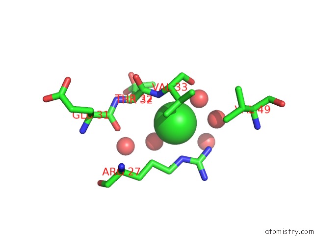
Mono view
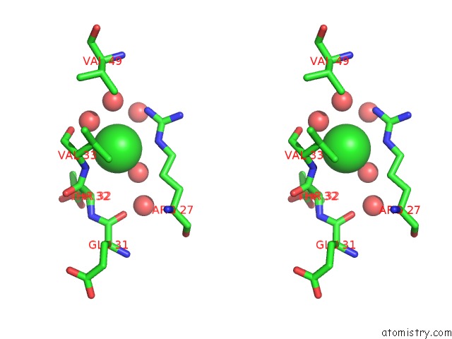
Stereo pair view
Mono view
Stereo pair view
A full contact list of Chlorine with other atoms in the Cl binding
site number 1 of X-Ray Crystal Structure of the H141N Mutant of Perosamine N- Acetyltransferase From Caulobacter Crescentus in Complex with Coa and Gdp-Perosamine within 5.0Å range:
|
Reference:
J.B.Thoden,
L.A.Reinhardt,
P.D.Cook,
P.Menden,
W.W.Cleland,
H.M.Holden.
Catalytic Mechanism of Perosamine N-Acetyltransferase Revealed By High-Resolution X-Ray Crystallographic Studies and Kinetic Analyses. Biochemistry V. 51 3433 2012.
ISSN: ISSN 0006-2960
PubMed: 22443398
DOI: 10.1021/BI300197H
Page generated: Sun Jul 21 12:41:40 2024
ISSN: ISSN 0006-2960
PubMed: 22443398
DOI: 10.1021/BI300197H
Last articles
Zn in 9J0NZn in 9J0O
Zn in 9J0P
Zn in 9FJX
Zn in 9EKB
Zn in 9C0F
Zn in 9CAH
Zn in 9CH0
Zn in 9CH3
Zn in 9CH1